Connect with us

Brain Injury

Capturing the Chemical Code: Scientists Stop ‘Brain Channels in Time’ to Reveal Learning Mechanism

The finding of a new study could advance the development of new drugs that block or open such signaling channels to treat conditions as varied as epilepsy and some intellectual disorders.
Avatar photo

Published

on

The article you provided is well-researched and informative, but I’ve rewritten it to improve clarity, structure, and style for a general audience. Here’s the rewritten content:

Capturing the Chemical Code: Scientists Stop ‘Brain Channels in Time’ to Reveal Learning Mechanism

Scientists have made a groundbreaking discovery about how brain cells communicate with each other. By using a highly specialized microscope, researchers were able to capture precise details of how one of the most common signaling molecules, glutamate, opens a channel and allows a flood of charged particles to enter. This finding could lead to the development of new treatments for conditions such as epilepsy and intellectual disorders.

The study, led by Johns Hopkins Medicine researchers, was published in the journal Nature. “Neurons are the cellular foundation of the brain, and the ability to experience our environment and learn depends on chemical communications between neurons,” explains Edward Twomey, Ph.D., assistant professor of biophysics and biophysical chemistry at the Johns Hopkins University School of Medicine.

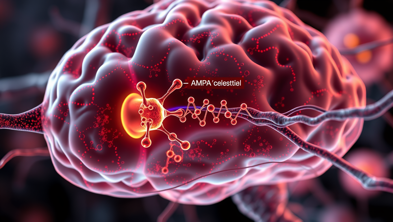
Scientists have long known that glutamate is a major molecule responsible for neuron-to-neuron communication. When glutamate binds to an AMPA receptor channel, it acts like a key that unlocks the door to the channel, enabling it to open more widely. This process creates electrical signals that form communications between neurons.

To understand this process in detail, researchers used a cryo-electron microscope (cryo-EM) to image these channels during specific steps in the communication processes. They purified AMPA receptors from lab-grown human embryonic cells and heated them to body temperature before exposing them to glutamate. Immediately after this, the receptors were flash frozen and analyzed with cryo-EM to get a snapshot of the AMPA receptors bound to glutamate.

After assembling over a million images taken with cryo-EM, the team found that glutamate molecules act like a key that unlocks the door to the channel, enabling it to open more widely. This occurs by the clamshell-like structure of the AMPA receptor closing around glutamate, an action that pulls open the channel below.

Twomey’s previous research has shown that drugs such as perampanel, used to treat epilepsy, act as a door stopper around the AMPA receptor to limit the channel from opening and reducing the abundance of activity known to happen in brain cells of people with epilepsy.

The findings could be used to develop new drugs that bind to AMPA receptors in different ways that either open or close the signaling channels of brain cells. “With each new finding, we are figuring out each of the building blocks that enable our brains to function,” says Twomey.

Additional scientists who contributed to the work are Anish Kumar Mondal from Johns Hopkins and Elisa Carrillo and Vasanthi Jayaraman from UTHealth Houston.

Funding for the research was provided by the National Institutes of Health (R35GM154904, R35GM122528), the Searle Scholars Program and the Diana Helis Henry Medical Research Foundation.

Biochemistry

A Breakthrough in Brain Research: The Iontronic Pipette Revolutionizes Neurological Studies

Researchers have developed a new type of pipette that can deliver ions to individual neurons without affecting the sensitive extracellular milieu. Controlling the concentration of different ions can provide important insights into how individual brain cells are affected, and how cells work together. The pipette could also be used for treatments.

Avatar photo

Published

on

The development of an iontronic pipette at Linköping University has opened up new avenues for neurological research. This innovative tool allows researchers to deliver ions directly to individual neurons without affecting the surrounding extracellular milieu. By controlling the concentration of various ions, scientists can gain valuable insights into how brain cells respond to different stimuli and interact with each other.

The human brain consists of approximately 85-100 billion neurons, supported by a similar number of glial cells that provide essential functions such as nutrition, oxygenation, and healing. The extracellular milieu, a fluid-filled space between the cells, plays a crucial role in maintaining cell function. Changes in ion concentration within this environment can activate or inhibit neuronal activity, making it essential to study how local changes affect individual brain cells.

Previous attempts to manipulate the extracellular environment involved pumping liquid into the area, disrupting the delicate biochemical balance and making it difficult to determine whether the substances themselves or the changed pressure were responsible for the observed effects. To overcome this challenge, researchers at the Laboratory of Organic Electronics developed an iontronic micropipette measuring only 2 micrometers in diameter.

This tiny pipette can deliver ions such as potassium and sodium directly into the extracellular milieu, allowing scientists to study how individual neurons respond to these changes. Glial cell activity is also monitored, providing a more comprehensive understanding of brain function.

Theresia Arbring Sjöström, an assistant professor at LOE, highlighted that glial cells are critical components of the brain’s chemical environment and can be precisely activated using this technology. In experiments conducted on mouse hippocampus tissue slices, it was observed that neurons responded dynamically to changes in ion concentration only after glial cell activity had saturated.

This research has significant implications for neurological disease treatment. The iontronic pipette could potentially be used to develop extremely precise treatments for conditions such as epilepsy, where brain function can be disrupted by localized imbalances in ion concentrations.

Researchers are now continuing their studies on chemical signaling in healthy and diseased brain tissue using the iontronic pipette. They also aim to adapt this technology to deliver medical drugs directly to affected areas of the brain, paving the way for more targeted treatments for neurological disorders.

Continue Reading

Brain Injury

“Resonating with Sound: Study Suggests Our Brains Physically Respond to Music”

Psychologists suggest our brains and bodies don’t just understand music, they physically resonate with it. These discoveries, based on findings in neuroscience, music, and psychology, support Neural Resonance Theory (NRT).

Avatar photo

Published

on

By

A groundbreaking study co-authored by McGill psychologist Caroline Palmer has revealed that our brains and bodies don’t just understand music; they physically resonate with it. This revolutionary discovery is based on findings in neuroscience, music, and psychology, supporting the Neural Resonance Theory (NRT).

According to NRT, musical experiences arise from the brain’s natural oscillations that sync with rhythm, melody, and harmony. This resonance shapes our sense of timing, musical pleasure, and the instinct to move with the beat.

“This theory suggests that music is powerful not just because we hear it, but because our brains and bodies become it,” said Palmer, Professor in the Department of Psychology at McGill and Director of the Sequence Production Lab. “That has big implications for therapy, education, and technology.”

The study’s publication in Nature Reviews Neuroscience marks the first time the entire NRT is being published in a single paper, she added.

NRT suggests that structures like pulse and harmony reflect stable resonant patterns in the brain, shared across people independent of their musical background. This theory explains how we hear and produce music through fundamental dynamical principles of human brain mechanisms that apply from the ear to the spinal cord and limb movements.

The study’s findings have significant potential applications:

Therapeutic tools for conditions like stroke, Parkinson’s, and depression

Emotionally intelligent AI that can respond to or generate music more like humans

New learning technologies to support rhythm and pitch education

Cross-cultural insight into why music connects people around the world

The study was led by Edward Large (University of Connecticut) and co-authored by Caroline Palmer. The research received funding from a Canada Research Chair and a NSERC Discovery Grant.

Continue Reading

Behavior

The Adaptive Nature of Object Recognition: How Experience Shapes Our Perception of Reality

New findings suggest neurons have much more functional dexterity than scientists previously realized.

Avatar photo

Published

on

By

The human brain has an incredible ability to recognize objects from a very young age. This process involves the visual cortex, which is responsible for processing visual information from the eyes. While it’s been thought that specific neurons along this pathway handle specific types of information depending on their location, new research suggests that feedback connections play a crucial role in object recognition. These connections convey information from higher cortical areas to lower ones, contributing to the dynamic capabilities of the brain.

Studies have shown that even at the first stages of object perception, neurons are sensitive to much more complex visual stimuli than previously believed. This capability is informed by feedback from higher cortical areas, which can adapt moment-to-moment to the information they’re receiving. In fact, researchers have found that a single neuron may be more responsive to one target and with another cue, they’ll be more responsive to a different target.

This adaptive processing allows the brain to dynamically tune its functional properties, changing its specificities with varying sensory experience. The findings of this research have significant implications for our understanding of how we perceive reality and could lead to new insights into the mechanisms underlying brain disorders such as autism.

In fact, researchers are now beginning to investigate animal models of autism at both the behavioral and imaging level. By studying perceptual differences between autism-model mice and their wild-type littermates, scientists hope to identify any cortical circuitry differences that may underlie these differences.

Overall, this research highlights the complex and dynamic nature of object recognition processes in the brain, and how experience shapes our perception of reality. It also underscores the importance of feedback connections in this process and has significant implications for our understanding of brain function and disorders.

Continue Reading

Trending