Connect with us
We’re experimenting with AI-generated content to help deliver information faster and more efficiently.
While we try to keep things accurate, this content is part of an ongoing experiment and may not always be reliable.
Please double-check important details — we’re not responsible for how the information is used.

Animal Learning and Intelligence

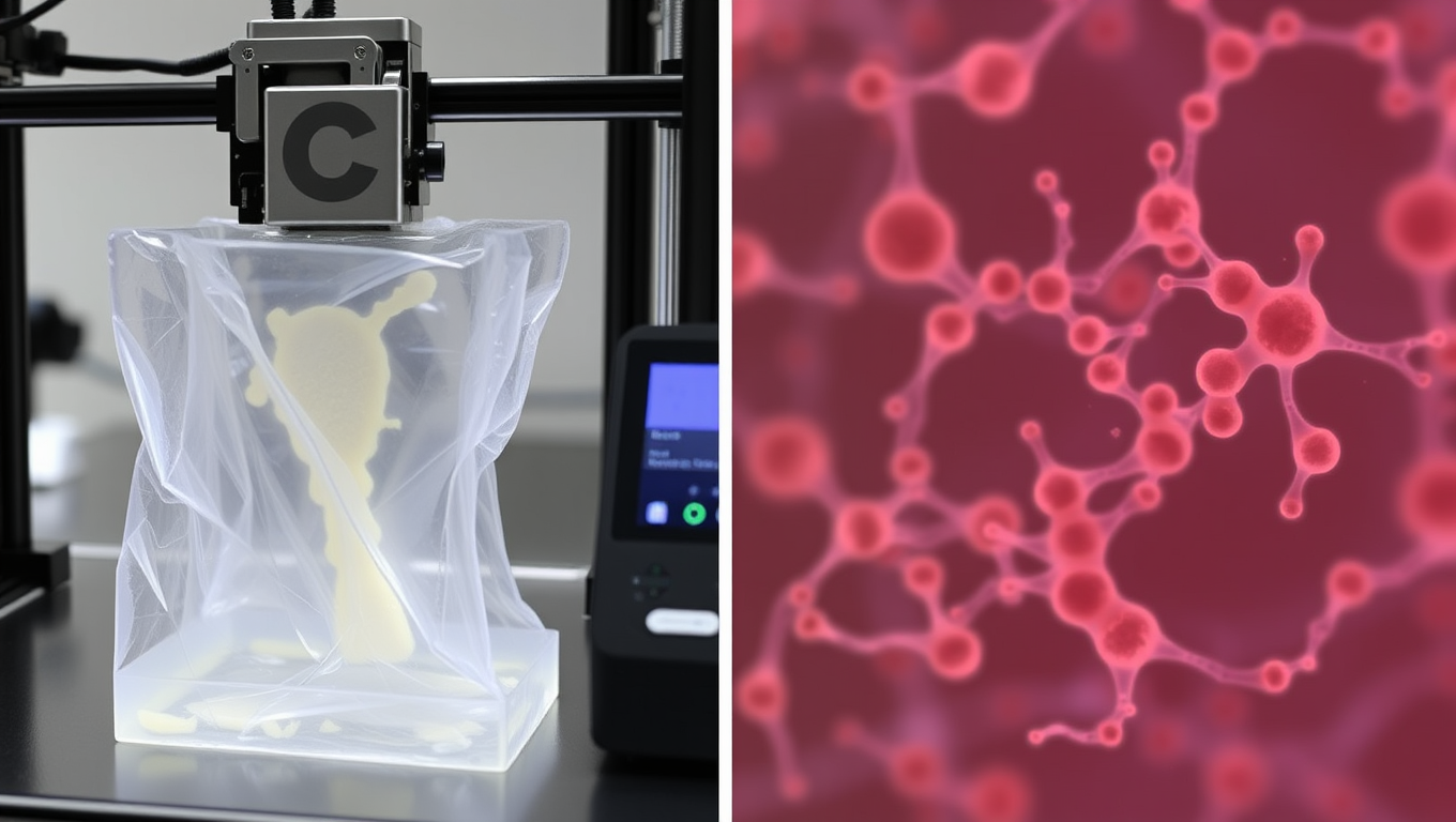
3D-Printed Skin Imitation Revolutionizes Cosmetics Testing

A research team is developing a 3D-printed skin imitation equipped with living cells in order to test nanoparticles from cosmetics without animal testing.

Avatar photo

Published

on

A groundbreaking research collaboration between TU Graz and the Vellore Institute of Technology in India is developing a revolutionary 3D-printed skin imitation that can replace animal testing for cosmetics. This innovative technology has the potential to transform the way we test the absorption and toxicity of nanoparticles from cosmetics, such as sun creams.

In response to Directive 2010/63/EU’s restrictions on animal testing within the EU, researchers are intensifying their search for alternatives. A team at TU Graz and VIT is working on creating skin imitations that mimic the native three-layer tissue structure and biomechanics of human skin using 3D printing.

The key to this breakthrough lies in developing hydrogel formulations that can interact with living skin cells. These hydrogels must not only support cell survival but also enable cell growth and multiplication. The researchers have successfully created stable and 3D-printable structures, which are then cross-linked for mechanical and chemical stabilisation.

TU Graz’s expertise in cross-linking methods ensures that the process takes place under mild conditions without using cytotoxic chemicals. Once the hydrogels are stabilised, they undergo rigorous testing at VIT to assess their resistance and toxicity in cell culture. Only when skin cells within the hydrogel survive for two to three weeks and develop into skin tissue can the researchers speak of a true skin imitation.

The first tests have been remarkably successful, with the 3D-printed hydrogels proving non-cytotoxic and mechanically stable. The next step will be using these skin imitations to test nanoparticles from cosmetics, marking a significant milestone in this collaborative research project.

Karin Stana Kleinschek from TU Graz notes that the complementary expertise between the two research institutions has been instrumental in achieving this success. By combining their strengths in material research and molecular cell biology, they are now working together to further optimise the hydrogel formulations and validate their usefulness as a substitute for animal experiments.

This 3D-printed skin imitation has the potential to revolutionise the way we test cosmetics, making it possible to create safer products without relying on animal testing. As researchers continue to refine this technology, they are one step closer to achieving a more humane and effective approach to product development.

Animal Learning and Intelligence

The Buzz on Caffeine: How it Affects the Sleeping Brain

Coffee can help you stay awake. But what does caffeine actually do to your brain once you’re asleep? Using AI, a team of researchers has an answer: it affects the brain’s ‘criticality’.

Avatar photo

Published

on

The article delves into the intricate relationship between caffeine and the sleeping brain, offering fresh insights from a recent study published in Nature Communications Biology. Researchers from Université de Montréal have shed new light on how caffeine can modify sleep patterns and influence the brain’s recovery during the night.

Led by Philipp Thölke, a research trainee at UdeM’s Cognitive and Computational Neuroscience Laboratory (CoCo Lab), the team used AI and electroencephalography (EEG) to study caffeine’s effects on sleep. Their findings reveal that caffeine increases the complexity of brain signals and enhances brain “criticality” during sleep – a state characterized by balanced order and chaos.

Interestingly, this effect is more pronounced in younger adults, particularly during REM sleep, the phase associated with dreaming. The researchers attribute this finding to a higher density of adenosine receptors in young brains, which naturally decrease with age. Adenosine is a molecule that accumulates throughout the day, causing fatigue.

The study’s lead author, Thölke, notes that caffeine stimulates the brain and pushes it into a state of criticality, where it is more awake, alert, and reactive. However, this state can interfere with rest at night, preventing the brain from relaxing or recovering properly.

The researchers used EEG to record the nocturnal brain activity of 40 healthy adults on two separate nights: one when they consumed caffeine capsules three hours before bedtime and another when they took a placebo at the same time. They applied advanced statistical analysis and artificial intelligence to identify subtle changes in neuronal activity, revealing that caffeine increased the complexity of brain signals during sleep.

The team also discovered striking changes in the brain’s electrical rhythms during sleep: caffeine attenuated slower oscillations such as theta and alpha waves – generally associated with deep, restorative sleep – and stimulated beta wave activity, which is more common during wakefulness and mental engagement.

These findings suggest that even during sleep, the brain remains in a more activated, less restorative state under the influence of caffeine. This change in the brain’s rhythmic activity may help explain why caffeine affects the efficiency with which the brain recovers during the night, with potential consequences for memory processing.

The study’s implications are significant, particularly given the widespread use of caffeine as a daily remedy for fatigue. The researchers stress the importance of understanding its complex effects on brain activity across different age groups and health conditions. They add that further research is needed to clarify how these neural changes affect cognitive health and daily functioning, potentially guiding personalized recommendations for caffeine intake.

Continue Reading

Animal Learning and Intelligence

The Brainy Advantage: How Asian Elephants Outshine Their African Relatives

African elephants are the largest land animals on earth and significantly larger than their relatives in Asia, from which they are separated by millions of years of evolution. Nevertheless, Asian elephants have a 20 percent heavier brain, as scientists were able to demonstrate. They also showed that elephant brains triple in weight after birth. These results provide potential explanations for behavioral differences between African and Asian elephants as well as for the pachyderms’ long youth, during which they gain enormous experience and learn social skills.

Avatar photo

Published

on

The article delves into the fascinating world of elephant brains, highlighting the distinct differences between their Asian and African counterparts. Despite being separated by millions of years of evolution, research has revealed that Asian elephants possess a 20% heavier brain than their larger African relatives. This groundbreaking finding, published in the scientific journal “PNAS Nexus,” sheds light on potential explanations for behavioral differences between the two species, as well as their remarkable youth and long lifespan.

Elephants are renowned for their exceptional social and intelligent nature, yet surprisingly little is known about their brains. A team of international researchers, led by Malav Shah and Michael Brecht from Humboldt-Universität zu Berlin, has analyzed the weight and structure of Asian elephant (Elephas maximus) and African elephant (Loxodonta africana) brains based on dissections of wild and zoo animals, as well as literature data and MRI scans. Their findings show that adult female Asian elephants have significantly heavier brains, weighing an average of 5,300 grams, compared to their African counterparts, which weigh around 4,400 grams.

Moreover, the cerebellum is proportionally heavier in African elephants (22% of total brain weight) than in Asian elephants (19%). The researchers attribute this difference to the more complex motor function of the trunk in African elephants, which can perform diverse movements with their two trunk fingers. This is also reflected in a higher number of neurons in the trunk’s control center in the brain.

The study further reveals that elephant brains grow almost as much as human brains after birth, tripling in weight by adulthood. This remarkable postnatal brain growth exceeds that of all primates, except humans, where the brain at birth weighs only around a fifth of its final weight. The researchers emphasize that this finding is significant for understanding motor skills and social behavior in elephants.

The study’s authors highlight the challenges involved in acquiring elephant brains for research, as extracting them from skulls is a complex veterinary procedure rarely performed. Nevertheless, they were able to analyze 19 brains extracted from deceased zoo animals or wild elephants, including those obtained from dissections of wild elephants that had died. The inclusion of data from an earlier study by another research team further strengthened their analysis.

The implications of these findings are profound, suggesting that the difference in brain weight could explain important behavioral differences between Asian and African elephants. For instance, while both species interact with humans differently, Asian elephants have been partially domesticated over thousands of years and are used as work animals in various cultures and regions. In contrast, there are only a few cases where domestication was even partially successful for African elephants.

The study’s authors conclude that social factors and learning processes could explain the strong brain growth after birth, as elephants live in complex social structures and have an outstanding memory. The experience and accumulated knowledge of adult elephants, especially matriarchs, is central to group behavior in elephants and young animals are closely cared for over a long period of childhood and adolescence.

Ultimately, this research highlights the need for further investigation into the brains of Asian and African elephants and their significance for motor skills and social behavior. As the authors note, there are many unanswered questions in researching these fascinating, intelligent animals and their “control centers.”

Continue Reading

Animal Learning and Intelligence

Mice Use Chemical Cues to Sense Social Hierarchy: A Fascinating Phenomenon Shared with Humans

Researchers have shown that mice use chemical cues, including odors, to detect the social rank of an unfamiliar mouse and compare it to their own, using this information to determine their behavior.

Avatar photo

Published

on

The researchers at the Francis Crick Institute have made a groundbreaking discovery: mice use chemical cues, including odors, to sense the social hierarchy of unfamiliar mice and compare it to their own. This remarkable phenomenon has been shown to influence the behavior of mice in confrontations with other mice.

Unlike previous suggestions that mice may display fixed behavior regardless of who they interact with, or that physical properties can give cues about social ranking, the new research published in Current Biology reveals that mice instead infer an unfamiliar mouse’s rank through chemical cues transmitted in the air (odors) or through direct contact (non-volatile scent cues).

The Crick team conducted a series of experiments to demonstrate this remarkable ability. They created a test where male mice entered a transparent tube at opposite ends, meeting in the middle. In this type of confrontation, a more submissive animal will typically retreat. The researchers first observed interactions between mice who shared the same cage, ranking each mouse on a hierarchy before observing how the mice responded to a set of unfamiliar opponents.

The results showed that the strangers could recognize each other’s rank, compare it to their own, and either retreat or force the other mouse to retreat. To further investigate this phenomenon, the team tested the mice in the dark, finding that this did not affect rank recognition, suggesting that cues like physical size or behavior don’t determine recognition of a more aggressive opponent.

The researchers then experimentally blocked the two chemosensory systems that mice use – one for odors in the air (olfactory system) and one for chemical signals transmitted by physical contact (vomeronasal system). They found no effect when just one of these systems was removed; both needed to be ablated before the mice couldn’t recognize opponent rank. This showed that mice use both olfactory and vomeronasal systems to recognize rank and can compensate if one is missing.

Like humans, mice are able to infer the social status of others around them relative to their own, using sensory cues such as language, facial expression, or clothing. The next step for the researchers is to investigate which areas of the brain process the information on opponent rank and own rank and initiate a decision to retreat or advance.

This remarkable phenomenon offers an interesting perspective on social mobility: humans, like mice, can enter a new group of people but still maintain understanding of our own social rank and gauge the social status of unfamiliar people. The State-Dependent Neural Processing Laboratory studies how processes within the brain are impacted by the state of the body, with the hope of advancing a more integrative view of brain physiology in health and disease.

Continue Reading

Trending