Connect with us
We’re experimenting with AI-generated content to help deliver information faster and more efficiently.
While we try to keep things accurate, this content is part of an ongoing experiment and may not always be reliable.
Please double-check important details — we’re not responsible for how the information is used.

Alzheimer's

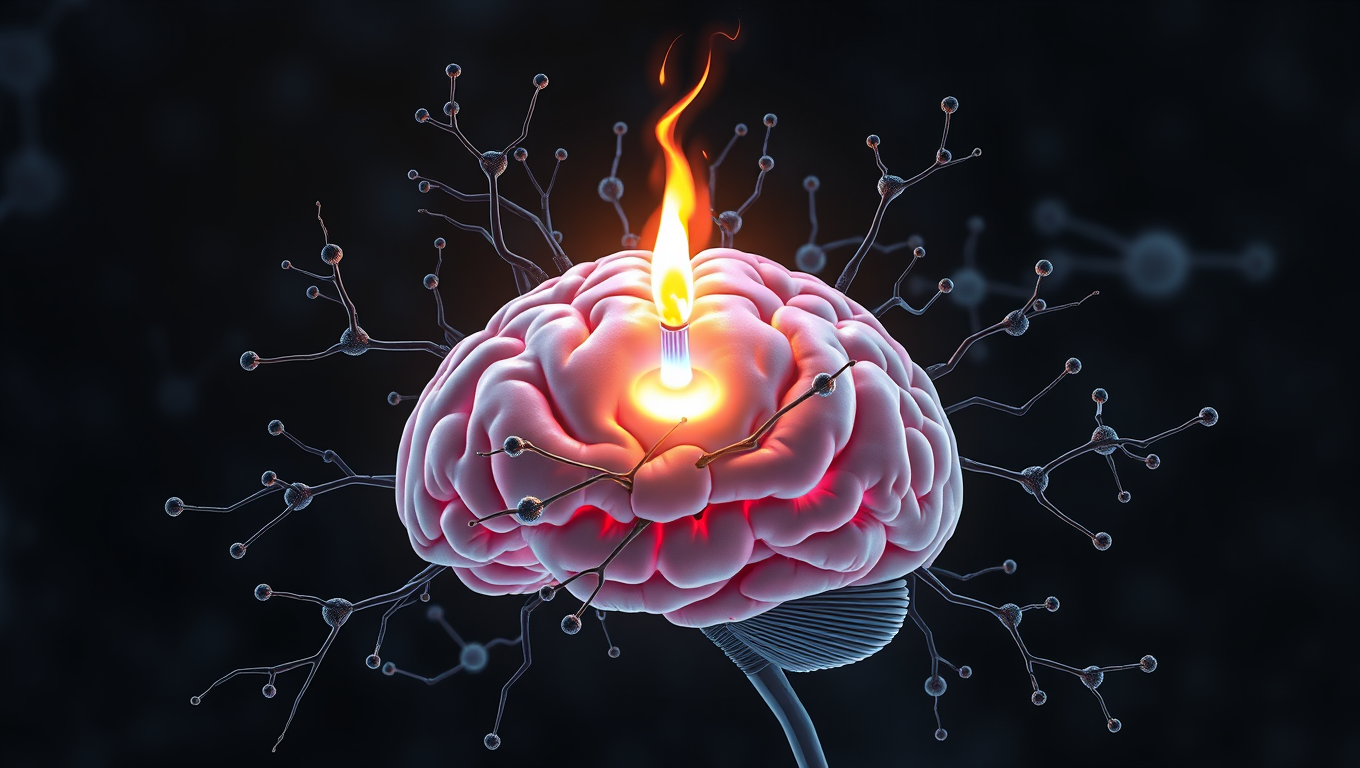
Breaking Down Brain Inflammation: Scientists Identify Key Enzyme in Alzheimer’s Disease

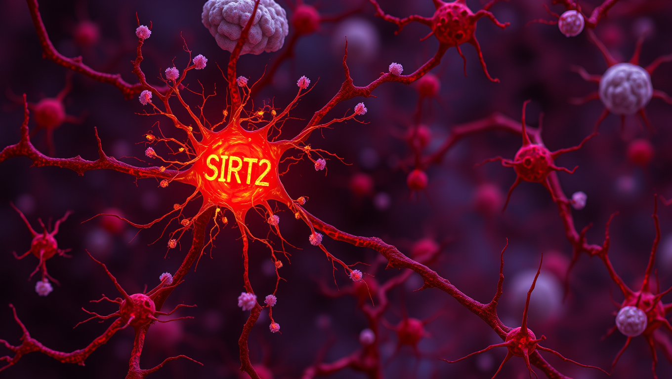
A research team has identified a previously unknown enzyme, SIRT2, that plays a key role in memory loss associated with Alzheimer’s disease (AD). The study provides critical insights into how astrocytes contribute to cognitive decline by producing excessive amounts of the inhibitory neurotransmitter GABA.
Avatar photo

Published

on

As researchers continue to unravel the mysteries of Alzheimer’s disease (AD), a team at the Institute for Basic Science (IBS) has made a significant breakthrough. They have identified a previously unknown enzyme, SIRT2, that plays a crucial role in memory loss associated with AD. This discovery provides critical insights into how astrocytes contribute to cognitive decline by producing excessive amounts of the inhibitory neurotransmitter GABA.

Astrocytes, once thought to only support neurons, are now known to actively influence brain function. In AD, astrocytes become reactive, attempting to clear amyloid-beta (Aβ) plaques, a hallmark of the disease. However, this process triggers a harmful chain reaction, leading to the overproduction of GABA, which dampens brain activity and causes memory impairment.

The IBS research team used molecular analysis, microscopic imaging, and electrophysiology to identify SIRT2 as one of the critical enzymes involved in GABA overproduction in AD-affected astrocytes. They found that SIRT2 protein was increased in the astrocytes of a commonly used AD mouse model and in post-mortem human AD patient brains.

“When we inhibited the astrocytic expression of SIRT2 in AD mice, we observed partial recovery of memory and reduced GABA production,” said Mridula Bhalla, the lead author of the study. “While we expected reduced GABA release, we found that only short-term working memory was recovered, and spatial memory was not. This was exciting but also left us with more questions.”
The research team’s findings suggest that SIRT2 participates in the last step of GABA production, while hydrogen peroxide (H2O2) is produced earlier in the process. They noted that inhibition of SIRT2 continued H2O2 production, indicating that neuronal degeneration might continue even though GABA production is reduced.

By identifying SIRT2 and ALDH1A1 as downstream targets, scientists can now selectively inhibit GABA production without affecting H2O2 levels. This breakthrough allows researchers to separate the effects of GABA and H2O2 and study their individual roles in neurodegeneration.

“This finding paves the way for more precise therapeutic strategies aimed at controlling astrocytic reactivity in Alzheimer’s disease,” said Director C Justin Lee. “We can now dissect the effects of GABA and H2O2 and study their individual roles in disease progression.” While SIRT2 may not be a direct drug target due to its limited effects on neurodegeneration, this research opens up new possibilities for treating AD.

Alzheimer's

Scientists Unlock Secret to Reversing Memory Loss by Boosting Brain’s Energy Engines

Scientists have discovered a direct cause-and-effect link between faulty mitochondria and the memory loss seen in neurodegenerative diseases. By creating a novel tool to boost mitochondrial activity in mouse models, researchers restored memory performance, suggesting mitochondria could be a powerful new target for treatments. The findings not only shed light on the early drivers of brain cell degeneration but also open possibilities for slowing or even preventing diseases like Alzheimer’s.

Avatar photo

Published

on

Scientists have made a groundbreaking discovery that could potentially reverse memory loss associated with neurodegenerative diseases. Researchers from Inserm and the University of Bordeaux, in collaboration with colleagues from the Université de Moncton in Canada, have successfully established a causal link between mitochondrial dysfunction and cognitive symptoms related to these conditions.

Mitochondria are tiny energy-producing structures within cells that provide the power needed for proper functioning. The brain is one of the most energy-demanding organs, relying on mitochondria to produce energy for neurons to communicate with each other. When mitochondrial activity is impaired, neurons fail to function correctly, leading to progressive neuronal degeneration and eventually, cell death.

In Alzheimer’s disease, for example, it has been observed that impaired mitochondrial activity precedes neuronal degeneration and ultimately, leads to memory loss. However, due to the lack of suitable tools, researchers were unable to determine whether mitochondrial alterations played a causal role in these conditions or were simply a consequence of the pathophysiological process.

In this pioneering study, researchers developed a unique tool that temporarily stimulates mitochondrial activity. By activating G proteins directly in mitochondria using an artificial receptor called mitoDreadd-Gs, they successfully restored both mitochondrial activity and memory performance in dementia mouse models.

“This work is the first to establish a cause-and-effect link between mitochondrial dysfunction and symptoms related to neurodegenerative diseases,” explains Giovanni Marsicano, Inserm research director. “Impaired mitochondrial activity could be at the origin of the onset of neuronal degeneration.”

The tool developed by researchers has opened doors to considering mitochondria as a new therapeutic target for treating memory loss associated with neurodegenerative diseases. Further studies are needed to measure the effects of continuous stimulation of mitochondrial activity and determine its potential impact on symptoms and neuronal loss.

Ultimately, this research holds promise for identifying molecular and cellular mechanisms responsible for dementia, facilitating the development of effective therapeutic targets, and potentially delaying or even preventing memory loss associated with neurodegenerative diseases.

Continue Reading

Alzheimer's

A Breakthrough in Brain Research: Scientists Grow a Mini Human Brain that Lights Up and Connects Like the Real Thing

Scientists at Johns Hopkins have grown a first-of-its-kind organoid mimicking an entire human brain, complete with rudimentary blood vessels and neural activity. This new “multi-region brain organoid” connects different brain parts, producing electrical signals and simulating early brain development. By watching these mini-brains evolve, researchers hope to uncover how conditions like autism or schizophrenia arise, and even test treatments in ways never before possible with animal models.

Avatar photo

Published

on

The groundbreaking research conducted at Johns Hopkins University has led to the creation of a novel whole-brain organoid, which is set to revolutionize the field of neuropsychiatric disorders. This miniature human brain, comprising neural tissues and rudimentary blood vessels, is an unprecedented achievement that could lead to a better understanding of conditions such as autism.

Lead author Annie Kathuria, an assistant professor in JHU’s Department of Biomedical Engineering, explained that most previous attempts at growing brain organoids focused on individual regions, such as the cortex or hindbrain. However, their research has succeeded in generating a multi-region brain organoid (MRBO), which represents a significant step forward.

The MRBO retains a broad range of types of neuronal cells, characteristic of a human brain in its early stages of development. This miniature brain weighs around 6 million to 7 million neurons compared to tens of billions found in adult brains. The researchers were able to stick the individual parts together using sticky proteins that act as a biological superglue.

As the tissues began to grow together, they started producing electrical activity and responding as a network. The creation of an early blood-brain barrier formation was also observed, which is essential for controlling molecule passage through the brain.

This breakthrough has far-reaching implications for studying neuropsychiatric disorders such as autism, schizophrenia, and Alzheimer’s disease. Whole-brain organoids will enable researchers to watch disorders develop in real-time, test experimental drugs, and tailor therapies to individual patients. The potential for improved clinical trial success is also substantial, with the current fail rate of 85% to 90% for neuropsychiatric drugs.

Using whole-brain organoids could lead to the discovery of new targets for drug screening and provide a more accurate representation of human brain development. As Kathuria emphasized, “We need to study models with human cells if you want to understand neurodevelopmental disorders or neuropsychiatric disorders.” The creation of this miniature human brain is an exciting step forward in brain research and has the potential to lead to significant advancements in the field.

Continue Reading

Alzheimer's

“Unlocking Brain Health: Scientists Discover Key Receptor for Microglia to Fight Alzheimer’s”

Scientists at UCSF have uncovered how certain immune cells in the brain, called microglia, can effectively digest toxic amyloid beta plaques that cause Alzheimer’s. They identified a key receptor, ADGRG1, that enables this protective action. When microglia lack this receptor, plaque builds up quickly, causing memory loss and brain damage. But when the receptor is present, it seems to help keep Alzheimer’s symptoms mild. Since ADGRG1 belongs to a drug-friendly family of receptors, this opens the door to future therapies that could enhance brain immunity and protect against Alzheimer’s in more people.

Avatar photo

Published

on

The scientific community has made significant strides in understanding the complex mechanisms behind Alzheimer’s disease. A recent study by researchers at UC San Francisco has shed light on how microglia, immune cells that play a crucial role in maintaining brain health, can break down and remove toxic proteins associated with the disease. This groundbreaking discovery could pave the way for novel therapeutic approaches to combat Alzheimer’s.

In Alzheimer’s, proteins like amyloid beta clump together, forming plaques that damage the brain. However, in some individuals, microglia effectively engulf and digest these proteins before they can cause harm. The resulting few and smaller clumps are associated with milder symptoms. Researchers at UCSF identified a molecular receptor, ADGRG1, which enables microglia to perform this critical function.

Using a mouse model of Alzheimer’s disease, the researchers observed that the loss of ADGRG1 led to a rapid buildup of amyloid plaques, neurodegeneration, and problems with learning and memory. The study also reanalyzed data from a prior human brain expression study, finding that individuals who died of mild Alzheimer’s had microglia with abundant ADGRG1, whereas those with severe Alzheimer’s had very little ADGRG1.

This discovery has significant implications for the development of new therapies. Since ADGRG1 is one of hundreds of G protein-coupled receptors targeted in drug development, it may be feasible to rapidly translate this finding into new treatments. As Dr. Piao noted, “Some people are lucky to have responsible microglia, but this discovery creates an opportunity to develop drugs to make microglia effective against amyloid-beta in everyone.” The potential for breakthrough therapies is exciting news for those affected by Alzheimer’s and their loved ones.

Continue Reading

Trending