
While we try to keep things accurate, this content is part of an ongoing experiment and may not always be reliable.
Please double-check important details — we’re not responsible for how the information is used.
Birth Defects
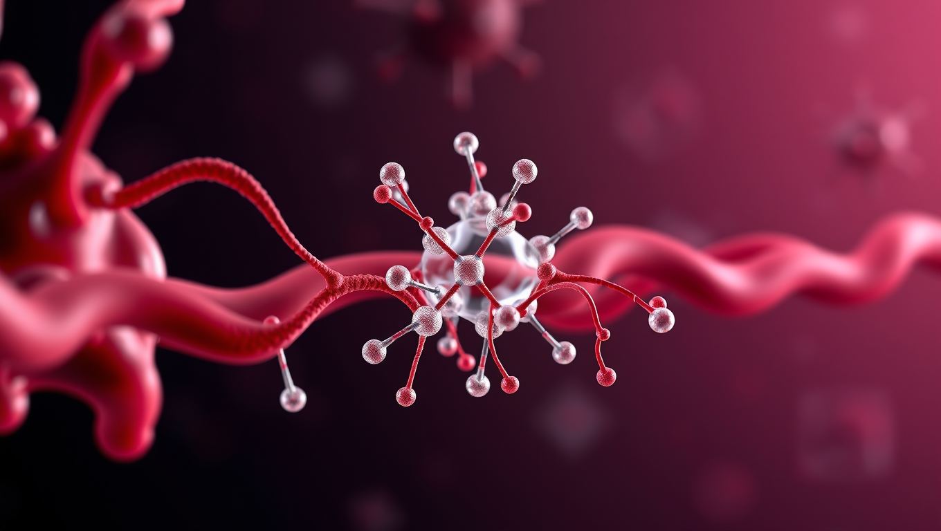
Breaking Down Barriers: Towards Gene-Targeting Drugs for Brain Diseases
Getting therapeutic drugs past the blood-brain barrier has long been a major challenge in treating brain diseases. Now, researchers have explored how cholesterol-modified heteroduplex oligonucleotides (Chol-HDOs) enhance drug delivery to the brain. Their study reveals that Chol-HDOs bind tightly to serum proteins, allowing them to persist in the bloodstream and cross into brain tissue. These findings offer insights into gene-targeting therapies and could help develop treatments for conditions like Alzheimer’s disease.


Autism
CRISPR-edited stem cells hold key to understanding autism spectrum disorder
A team at Kobe University has created a game-changing resource for autism research: 63 mouse embryonic stem cell lines, each carrying a genetic mutation strongly associated with the disorder. By pairing classic stem cell manipulation with precise CRISPR gene editing, they ve built a standardized platform that mirrors autism-linked genetic conditions in mice. These models not only replicate autism-related traits but also expose key dysfunctions, like the brain s inability to clean up faulty proteins.

Back and Neck Pain
Unveiling the Secrets of the Universe: The Largest-ever Map Reveals 10x More Early Galaxies Than Expected
An international team of scientists has unveiled the largest and most detailed map of the universe ever created using the James Webb Space Telescope, revealing nearly 800,000 galaxies stretching back to almost the beginning of time. The COSMOS-Web project not only challenges long-held beliefs about galaxy formation in the early universe but also unexpectedly revealed 10 times more galaxies than anticipated along with supermassive black holes Hubble couldn t see.

Autism
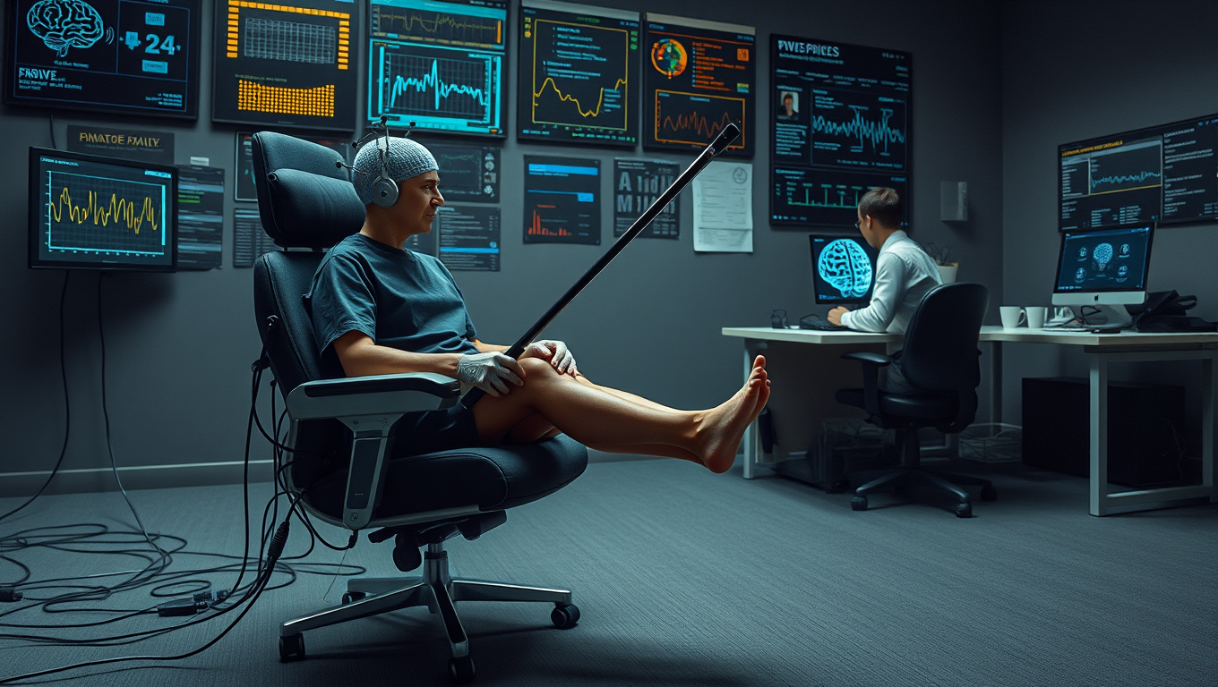
The Elusive Science of Tickling: Unraveling the Mysteries of a 2000-Year-Old Enigma
How come you can’t tickle yourself? And why can some people handle tickling perfectly fine while others scream their heads off? Neuroscientists argue that we should take tickle research more seriously.

A New Horizon for Vision: How Gold Nanoparticles May Restore People’s Sight

Revolutionizing Quantum Communication: Direct Connections Between Multiple Processors

Retiring Abroad Can Be Lonely Business
Harnessing Water Waves: A Breakthrough in Controlling Floating Objects

Household Electricity Three Times More Expensive Than Upcoming ‘Eco-Friendly’ Aviation E-Fuels, Study Reveals

“Unveiling Hidden Patterns: A New Twist on Interference Phenomena”
Reducing Falls Among Elderly Women with Polypharmacy through Exercise Intervention
-
 Detectors1 year ago
A New Horizon for Vision: How Gold Nanoparticles May Restore People’s Sight
-
Cancer1 year ago
Revolutionizing Quantum Communication: Direct Connections Between Multiple Processors
-
Earth & Climate1 year ago
Retiring Abroad Can Be Lonely Business
-
Albert Einstein1 year ago
Harnessing Water Waves: A Breakthrough in Controlling Floating Objects
-
Earth & Climate1 year ago
Household Electricity Three Times More Expensive Than Upcoming ‘Eco-Friendly’ Aviation E-Fuels, Study Reveals
-
Chemistry1 year ago
“Unveiling Hidden Patterns: A New Twist on Interference Phenomena”
-
Diseases and Conditions1 year ago
Reducing Falls Among Elderly Women with Polypharmacy through Exercise Intervention
-
Agriculture and Food1 year ago
“A Sustainable Solution: Researchers Create Hybrid Cheese with 25% Pea Protein”


