Connect with us
We’re experimenting with AI-generated content to help deliver information faster and more efficiently.
While we try to keep things accurate, this content is part of an ongoing experiment and may not always be reliable.
Please double-check important details — we’re not responsible for how the information is used.

Biotechnology

Unveiling the Origins of Gonadotrophs: A New Understanding of Puberty and Reproduction

Researchers have shown that gonadotrophs, cells in the pituitary gland with a key role in puberty and reproduction, come from two different populations, with the majority produced after birth rather than in the embryo, as previously thought.
Avatar photo

Published

on

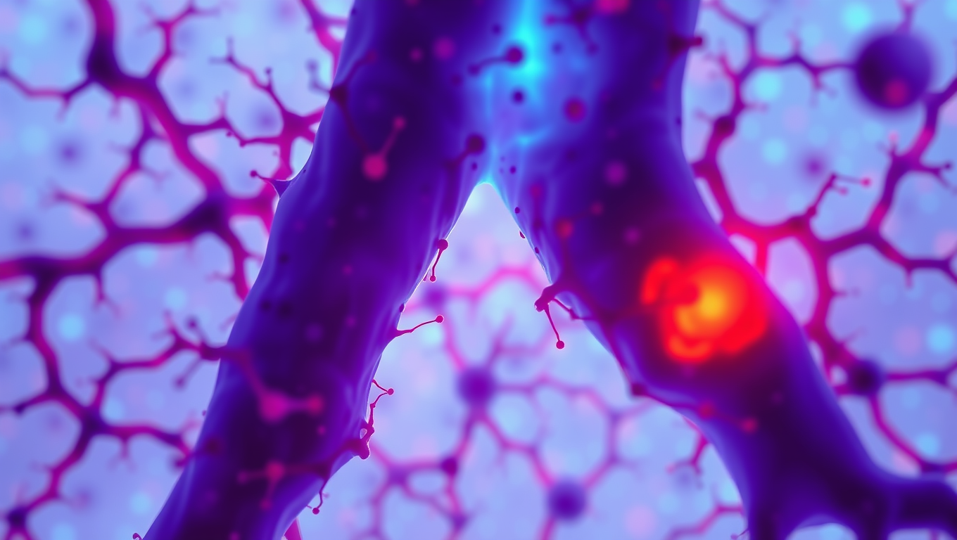
The Francis Crick Institute has made a groundbreaking discovery in the field of reproductive biology. Researchers have found that gonadotrophs, cells responsible for stimulating puberty and reproduction, originate from two distinct populations. This revelation challenges the long-held assumption that all gonadotrophs are produced during embryonic development.

A research team at the Crick Institute initially identified a population of tissue-specific stem cells in the pituitary gland. These stem cells were thought to be relatively insignificant, but further investigation revealed their crucial role in producing gonadotrophs. The team employed genetic marking and tracing techniques to follow the descendants of these stem cells as they developed into different cell types within the pituitary gland.

In mice studies, it was observed that the stem cell pool predominantly gave rise to gonadotrophs after birth, specifically during the “minipuberty” period. This process continued until puberty, highlighting a unique opportunity for intervention in disorders affecting reproduction. The team also discovered that the two populations of gonadotrophs are located in separate compartments within the pituitary gland and that the embryonic population remains stable throughout life.

The researchers further explored what stimulates these stem cells to become gonadotrophs specifically, confirming that it involves a physiological context rather than a specific hormone. They speculate that something about leaving the mother’s body at birth might be crucial for gonadotroph development at the right time.

This study has significant implications for understanding and treating disorders that impact puberty and fertility. The discovery of two distinct populations of gonadotrophs provides a new window of opportunity for diagnosis and intervention in conditions like congenital hypogonadotropic hypogonadism (CHH). By identifying this earlier, healthcare professionals can prevent children from failing to go through puberty later in life.

The lead researcher, Karine Rizzoti, emphasized the importance of this discovery: “We’ve known about this population of stem cells for a while, but it took the right tools used at the right time to see just how important they are. Instead of the previously held idea that gonadotrophs all have the same origin, we instead found that there are two waves of generation, before and after birth.”

This research has far-reaching implications for our understanding of reproductive biology and its potential applications in treating related disorders. The discovery of distinct populations of gonadotrophs opens up new avenues for investigation and highlights the importance of considering physiological context in the development of gonadotrophs.

Animals

Sugar-Based Sensors Revolutionize Snake Venom Detection

Researchers have published the first example of a synthetic sugar detection test for snake venom, offering a new route to rapid diagnosis and better antivenoms.

Avatar photo

Published

on

The detection of snake venom is a crucial step in treating life-threatening snake bites. According to the World Health Organization (WHO), every five minutes, 50 people are bitten by a snake worldwide, resulting in four permanent disabilities and one death. Traditional methods for diagnosing snake venom rely on antibodies, which have limitations such as high costs, lengthy procedures, and inconsistencies.

Researchers at the University of Warwick have made a groundbreaking discovery that could revolutionize snake venom detection. They have developed a glycopolymer-based ultraviolet-visible (UV-vis) test to detect Western Diamondback Rattlesnake (Crotalus atrox) venom. This new assay is a cheap and rapid alternative to antibody-based approaches, showcasing a version that specifically detects Crotalus atrox venom.

Dr. Alex Baker, lead researcher of the Baker Humanitarian Chemistry Group, explained that snake venoms are complex, making it challenging to detect toxins in the body. However, their research has produced an assay using synthetic sugars that mimic the natural sugar receptors targeted by venom proteins. The team engineered synthetic chains of sugar-like units (glycopolymers) attached to gold nanoparticles to amplify the response and make the reaction visible.

The Western Diamondback Rattlesnake venom binds to specific sugar molecules on red blood cells and platelets, disrupting blood clotting or interfering with immune responses leading to disability and death. The new assay changes color when venom toxins bind to the synthetic sugars, providing a rapid and cheap detection method beyond antibody-based techniques.

Mahdi Hezwani, first author of the research paper, emphasized that this assay could be a game-changer for snake envenomation. The team tested venom from other snake species, such as the Indian Cobra (Naja naja), and found that it did not interact with glycans in the body. This suggests that the new assay may have potential to distinguish between different snake venoms based on their sugar-binding properties.

This is the first example of a diagnosis test using sugars for detecting snake venom in a rapid detection system, building on the work of the Warwick research group using a glyconanoparticle platform in COVID-19 detection. The new assay is faster, cheaper, and easier to store, making it a more practical solution for treating snake bites.

The University of Warwick’s STEM Connect programme has enabled this innovative research, demonstrating the potential for bold and innovative solutions in addressing global health challenges.

Continue Reading

Biochemistry Research

“Unlocking Nature’s Math: Uncovering Gauge Freedoms in Biological Models”

Scientists have developed a unified theory for mathematical parameters known as gauge freedoms. Their new formulas will allow researchers to interpret research results much faster and with greater confidence. The development could prove fundamental for future efforts in agriculture, drug discovery, and beyond.

Avatar photo

Published

on

In the intricate language of mathematics, there lies a fascinating phenomenon known as gauge freedoms. This seemingly abstract concept may seem far removed from our everyday lives, but its impact is felt deeply in the realm of biological sciences. Researchers at Cold Spring Harbor Laboratory (CSHL) have made groundbreaking strides in understanding and harnessing this power.

Gauge freedoms are essentially the mathematical equivalent of having multiple ways to describe a single truth. In science, when modeling complex systems like DNA or protein sequences, different parameters can result in identical predictions. This phenomenon is crucial in fields like electromagnetism and quantum mechanics. However, until now, computational biologists have had to employ various ad hoc methods to account for gauge freedoms, rather than tackling them directly.

CSHL’s Associate Professor Justin Kinney, along with colleague David McCandlish, led a team that aimed to change this. They developed a unified theory for handling gauge freedoms in biological models. This breakthrough could revolutionize applications across multiple fields, from plant breeding to drug development.

Gauge freedoms are ubiquitous in computational biology, says Prof. Kinney. “Historically, they’ve been dealt with as annoying technicalities.” However, through their research, the team has shown that understanding and systematically addressing these freedoms can lead to more accurate and faster analysis of complex genetic datasets.

Their new mathematical theory provides efficient formulas for a wide range of biological applications. These formulas will empower scientists to interpret research results with greater confidence and speed. Furthermore, the researchers have published a companion paper revealing where gauge freedoms originate – in symmetries present within real biological sequences.

As Prof. McCandlish notes, “We prove that gauge freedoms are necessary to interpret the contributions of particular genetic sequences.” This finding underscores the significance of understanding gauge freedoms not just as a theoretical concept but also as a fundamental requirement for advancing future research in agriculture, drug discovery, and beyond.

This rewritten article aims to clarify complex scientific concepts for a broader audience while maintaining the original message’s integrity.

Continue Reading

Biology

Yeast Revolutionizes Human Protein Production with DNase1 Breakthrough

The protein DNase1 is one of the oldest biological agents in history: It has been on the market since 1958 and is now used, among other things, to treat cystic fibrosis. However, it takes considerable effort to produce it in immortalized hamster cells. This process is also costly. It would be far more cost-effective to produce it with undemanding yeast cells.

Avatar photo

Published

on

The production of human proteins has long been a costly and labor-intensive process, often relying on mammalian cells. However, a team led by Dr. Markus Napirei at Ruhr University Bochum has successfully produced the human protein DNase1 using yeast cells, a breakthrough that could revolutionize the field.

DNase1 is an enzyme used to treat cystic fibrosis and other conditions, but its production in mammalian cells has been limited by high costs and effort. The new method uses Pichia pastoris, a type of yeast fungus, to produce the protein, which can be stably integrated into the yeast genome and released as desired.

“This is the result of years of work, and could lay the groundwork for the manufacture of human DNase1 in yeast as a biological agent,” says Dr. Napirei. The research was published in PLOS ONE on April 29, 2025.

The advantages of using yeast cells over mammalian cells are clear: cost-effective culture conditions, high reproduction rates without the need to immortalize cells, and lower susceptibility to pathogens. In his doctoral thesis, Jan-Ole Krischek successfully expressed human DNase1 in Pichia pastoris, cleaned it, and characterized it for the first time.

One of the surprising findings was that the yeast produced considerably less human DNase1 than the mouse DNase1 used as a guide, despite sharing 82 percent of their primary structure. This is partly due to specific folding behaviors of the two proteins, explains Dr. Napirei.

DNase1 has been used for over 60 years to treat various conditions, including cystic fibrosis. The enzyme degrades cell-free DNA that can induce symptoms of illness. Inhaled DNase1 liquifies DNA-laden bronchial mucus, making it easier to cough up. Its potential use in other pathological processes is vast, particularly in the removal of neutrophil extracellular traps (NETs) and microthrombi that contain high levels of NET components.

Dr. Napirei suggests that DNase1 could be used to better dissolve microthrombi containing DNA, an application currently being explored in clinical studies. Another potential use is in dissolving thrombosis of a cerebral artery in the case of ischemic strokes.

This breakthrough has significant implications for the production and use of human proteins, particularly DNase1. The ability to produce this enzyme using yeast cells could lead to more cost-effective and efficient treatment options for patients, ultimately improving their quality of life.

Continue Reading

Trending