Connect with us
We’re experimenting with AI-generated content to help deliver information faster and more efficiently.
While we try to keep things accurate, this content is part of an ongoing experiment and may not always be reliable.
Please double-check important details — we’re not responsible for how the information is used.

Dementia

Alzheimer’s risk may start at the brain’s border, not inside it

Your brain has its own elite defense team — and new research shows these “guardian” cells might be the real battleground for neurological diseases like Alzheimer’s and stroke. Scientists discovered that most genetic risks linked to these diseases act not in neurons, but in the blood vessels and immune cells that form the blood-brain barrier.
Avatar photo

Published

on

Here is the rewritten article:

Alzheimer’s risk may start at the brain’s border, not inside it

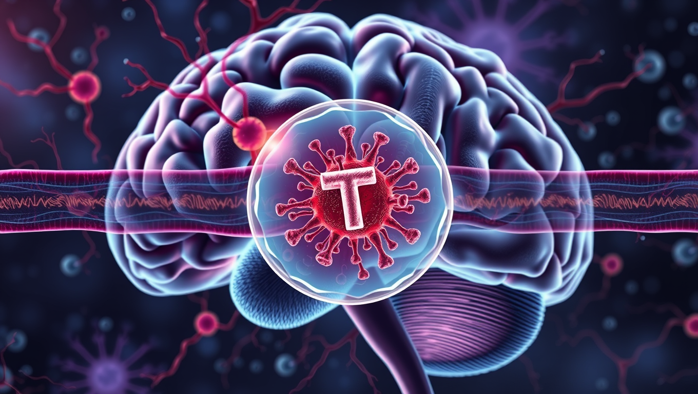
The brain’s health depends on more than just its neurons. A complex network of blood vessels and immune cells acts as the brain’s dedicated guardians – controlling what enters, cleaning up waste, and protecting it from threats by forming the blood-brain barrier.

A new study from Gladstone Institutes and UC San Francisco (UCSF) reveals that many genetic risk factors for neurological diseases like Alzheimer’s and stroke exert their effects within these very guardian cells.

“When studying diseases affecting the brain, most research has focused on its resident neurons,” says Gladstone Investigator Andrew C. Yang, PhD, senior author of the new study. “I hope our findings lead to more interest in the cells forming the brain’s borders, which might actually take center stage in diseases like Alzheimer’s.”
The findings, published in Neuron, address a long-standing question about where genetic risk begins and suggest that vulnerabilities in the brain’s defense system may be a key trigger for disease.

For years, large-scale genetic studies have linked dozens of DNA variants to a higher risk of neurological diseases like Alzheimer’s, Parkinson’s, or multiple sclerosis. Yet, a major mystery has persisted: over 90% of these variants lie not in the genes themselves, but in the surrounding DNA that does not contain the code for making proteins, once dismissed as “junk DNA.”

These regions act as complex dimmer switches, turning genes on or off. Until now, scientists haven’t had a full map of which switches control which genes or in which specific brain cells they operate, hindering the path from genetic discovery to new treatments.

A New Technology Finds Answers

The blood-brain barrier is the brain’s frontline defense – a cellular border made up of blood vessel cells, immune cells, and other supporting cells that meticulously controls access to the brain. Yet, these important cells have been difficult to study, even using the field’s most powerful genetic techniques.

To overcome this, the Gladstone team developed MultiVINE-seq, a technology that allows for the simultaneous analysis of multiple genomic and transcriptomic datasets from individual cells.

The new study utilized this technology to investigate the role of immune cells in the brain’s defense system. The researchers found that certain genetic variants associated with Alzheimer’s disease were more prevalent in immune cells than in neurons or other brain cells.

This suggests that the brain’s guardian cells may play a crucial role in the development and progression of Alzheimer’s disease.
The study also identified a potential new target for treating Alzheimer’s disease: PTK2B, a protein that is involved in the regulation of immune cell activity. The researchers found that therapies targeting PTK2B are already being developed for cancer treatment, and could potentially be repurposed for Alzheimer’s disease.

Location, Location, Location

The study’s findings on the brain’s “guardian” cells point to two new opportunities for protecting the brain. Located at the critical interface between the brain and the body, the cells are continually influenced by lifestyle and environmental exposures, which could synergize with genetic predispositions to drive disease.

Their location also makes them a promising target for future therapies, potentially allowing for drugs that can bolster the brain’s defenses from the “outside” without needing to cross the formidable blood-brain barrier. This work brings the brain’s vascular and immune cells into the spotlight, and could inform new, more accessible drug targets and lifestyle interventions to protect the brain from the outside in.

About the Study

The study, “Human brain vascular multi-omics elucidates disease risk associations,” was published in the journal Neuron on July 28, 2025. The work was supported by several funding agencies, including the National Institute of Neurological Disorders and Stroke, Alzheimer’s Association, BrightFocus Foundation, Cure Alzheimer’s Fund, Ludwig Family Foundation, Dolby Family Fund, Bakar Aging Research Institute, National Institute of Mental Health, National Institute of Aging, Leducq Foundation, and Joachim Herz Foundation.

Alzheimer's

“Unlocking Brain Health: Scientists Discover Key Receptor for Microglia to Fight Alzheimer’s”

Scientists at UCSF have uncovered how certain immune cells in the brain, called microglia, can effectively digest toxic amyloid beta plaques that cause Alzheimer’s. They identified a key receptor, ADGRG1, that enables this protective action. When microglia lack this receptor, plaque builds up quickly, causing memory loss and brain damage. But when the receptor is present, it seems to help keep Alzheimer’s symptoms mild. Since ADGRG1 belongs to a drug-friendly family of receptors, this opens the door to future therapies that could enhance brain immunity and protect against Alzheimer’s in more people.

Avatar photo

Published

on

The scientific community has made significant strides in understanding the complex mechanisms behind Alzheimer’s disease. A recent study by researchers at UC San Francisco has shed light on how microglia, immune cells that play a crucial role in maintaining brain health, can break down and remove toxic proteins associated with the disease. This groundbreaking discovery could pave the way for novel therapeutic approaches to combat Alzheimer’s.

In Alzheimer’s, proteins like amyloid beta clump together, forming plaques that damage the brain. However, in some individuals, microglia effectively engulf and digest these proteins before they can cause harm. The resulting few and smaller clumps are associated with milder symptoms. Researchers at UCSF identified a molecular receptor, ADGRG1, which enables microglia to perform this critical function.

Using a mouse model of Alzheimer’s disease, the researchers observed that the loss of ADGRG1 led to a rapid buildup of amyloid plaques, neurodegeneration, and problems with learning and memory. The study also reanalyzed data from a prior human brain expression study, finding that individuals who died of mild Alzheimer’s had microglia with abundant ADGRG1, whereas those with severe Alzheimer’s had very little ADGRG1.

This discovery has significant implications for the development of new therapies. Since ADGRG1 is one of hundreds of G protein-coupled receptors targeted in drug development, it may be feasible to rapidly translate this finding into new treatments. As Dr. Piao noted, “Some people are lucky to have responsible microglia, but this discovery creates an opportunity to develop drugs to make microglia effective against amyloid-beta in everyone.” The potential for breakthrough therapies is exciting news for those affected by Alzheimer’s and their loved ones.

Continue Reading

Alzheimer's

Uncovering the Hidden Culprits Behind Alzheimer’s Disease

A surprising new study has uncovered over 200 misfolded proteins in the brains of aging rats with cognitive decline, beyond the infamous amyloid and tau plaques long blamed for Alzheimer’s. These shape-shifting proteins don’t clump into visible plaques, making them harder to detect but potentially just as harmful. Scientists believe these “stealth” molecules could evade the brain’s cleanup systems and quietly impair memory and brain function. The discovery opens a new frontier in understanding dementia and could lead to entirely new targets for treatment and prevention.

Avatar photo

Published

on

Uncovering the Hidden Culprits Behind Alzheimer’s Disease

For decades, researchers have been trying to understand the root causes of Alzheimer’s disease. While amyloids, such as A-beta and tau proteins, have long been the focus of attention, a new study suggests that these sticky brain plaques may not be the only culprits behind cognitive decline.

Researchers at Johns Hopkins University have made a groundbreaking discovery, identifying over 200 types of misfolded proteins in rats that could contribute to age-related cognitive decline. This finding has significant implications for Alzheimer’s research and opens up new avenues for potential therapeutic targets and treatments.

“We’re seeing hundreds of proteins misfolding in ways that don’t clump together in an amyloid and yet still seem to impact how the brain functions,” said Stephen Fried, an assistant professor of chemistry and protein scientist. “Our research is showing that amyloids are just the tip of the iceberg.”

To reach this conclusion, Fried and his team studied 17 two-year-old rats with varying levels of cognitive impairment. They measured over 2,500 types of protein in the hippocampus, a part of the brain associated with spatial learning and memory. The researchers were able to determine which proteins misfolded for all the rats and are associated with aging in general versus which proteins specifically misfold in cognitively impaired rats.

More than 200 proteins were found to be misfolded in the cognitively impaired rats but maintained their shapes in the cognitively healthy rats. This suggests that some of these misfolded proteins may contribute to cognitive decline, according to the researchers.

Misfolded proteins are unable to carry out tasks necessary for a cell to function properly, so cells have a natural surveillance system that identifies and destroys these misbehaving proteins. However, it appears that some misfolded proteins can escape this surveillance system and still cause problems.

The next step for Fried’s team is to use high-resolution microscopes to get a more detailed picture of what the misfolded proteins look like at the molecular level.

“A lot of us have experienced a loved one or a relative who has become less capable of doing those everyday tasks that require cognitive abilities,” Fried said. “Understanding what’s physically going on in the brain could lead to better treatments and preventive measures.”

This research has significant implications for Alzheimer’s disease, as it suggests that there may be multiple targets for treatment beyond amyloids alone. By understanding the molecular differences between healthy and cognitively impaired brains, researchers can develop more effective treatments and potentially prevent cognitive decline in the first place.

Continue Reading

Alzheimer's

Uncovering the Hidden Defenses Against Alzheimer’s Disease: A Breakthrough Study on Brain Resilience

Scientists at UCSF combined advanced brain-network modeling, genetics, and imaging to reveal how tau protein travels through neural highways and how certain genes either accelerate its toxic journey or shield brain regions from damage. Their extended Network Diffusion Model pinpoints four gene categories that govern vulnerability or resilience, reshaping our view of Alzheimer’s progression and spotlighting fresh therapeutic targets.

Avatar photo

Published

on

Alzheimer’s disease is a complex condition that affects different parts of the brain in various ways. One key factor in the progression of the disease is the misbehavior of tau proteins, which can lead to toxic clumps forming inside neurons and impairing their function. Researchers have long sought to understand why some areas of the brain are more resilient to Alzheimer’s than others, a phenomenon known as selective vulnerability or resilience.

A recent study by researchers at the University of California, San Francisco (UCSF) has made significant strides in this area by combining advanced mathematical modeling with brain imaging and genetics. The study, published in Brain, identified multiple distinct pathways through which risk genes confer vulnerability or resilience to Alzheimer’s disease.

The researchers developed a model called the extended Network Diffusion Model (eNDM), which predicted where tau protein would spread next based on real-world brain connection data from healthy individuals. By applying this model to brain scans of 196 people at various stages of Alzheimer’s, they were able to identify areas that were resistant or vulnerable to the disease.

The study revealed four distinct types of genes: those that boost tau spread along the brain’s wiring (Network-Aligned Vulnerability), those that promote tau buildup in ways unrelated to connectivity (Network-Independent Vulnerability), those that help protect regions that are otherwise tau hotspots (Network-Aligned Resilience), and those that offer protection outside of the network’s usual path (Network-Independent Resilience).

These findings have significant implications for understanding Alzheimer’s disease and developing potential intervention targets. The study’s lead author, Ashish Raj, PhD, noted that their research offers a “hopeful map forward” in understanding and eventually stopping Alzheimer’s disease.

The researchers also highlighted the importance of considering the different biological functions of genes that respond independently of the network versus those that respond in concert with it. This nuanced approach could lead to more effective strategies for identifying potential intervention targets and developing treatments for Alzheimer’s disease.

Continue Reading

Trending