Connect with us

Biochemistry

Democratizing Chemical Analysis with Machine Learning and Robotics

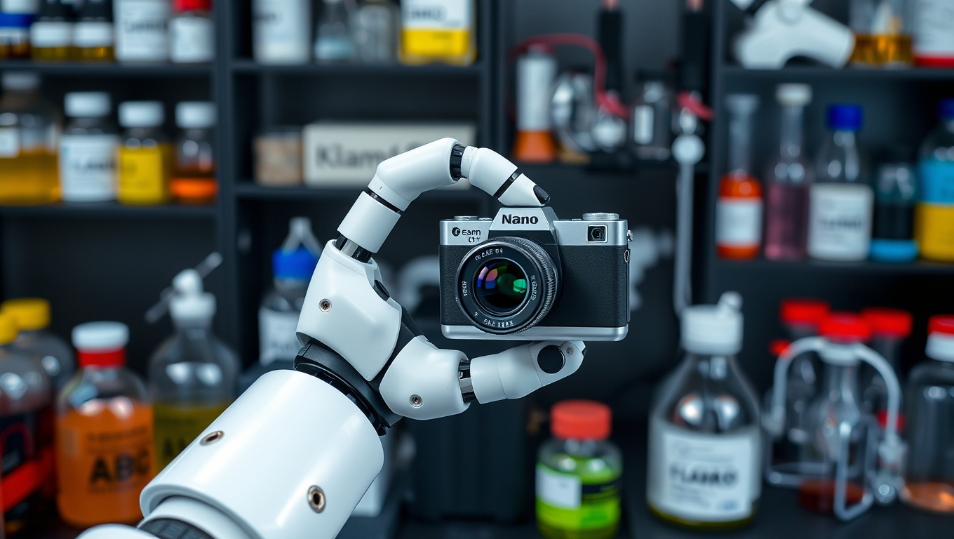
Chemists have created a machine learning tool that can identify the chemical composition of dried salt solutions from an image with 99% accuracy. By using robotics to prepare thousands of samples and artificial intelligence to analyze their data, they created a simple, inexpensive tool that could expand possibilities for performing chemical analysis.
Avatar photo

Published

on

Democratizing Chemical Analysis with Machine Learning and Robotics

A team of chemists at Florida State University has made significant strides in developing a machine learning tool that can identify the chemical composition of dried salt solutions from an image with remarkable accuracy. This breakthrough, published in Digital Discovery, has far-reaching implications for various fields, including space exploration, law enforcement, home testing, and more.

“We are living in the age of artificial intelligence and big data,” said co-author Oliver Steinbock, a professor in the FSU Department of Chemistry and Biochemistry. “We thought that if we used sufficiently large databases with enough pictures of different compounds and stains, we could maybe use AI to determine what the composition is.”
The research built upon a previous study from Steinbock’s lab, where researchers used machine learning to identify the chemical composition of salt stains from photos. In this new paper, they amplified that work by using a robot to process samples that were later analyzed by an improved machine learning program.

The Robotic Drop Imager (RODI) was created to prepare more than 2,000 samples per day, allowing the researchers to build a library of over 23,000 images. After preparing samples and taking photos, they simplified each image by converting them to grayscale and extracted 47 features, such as pattern area, brightness, and other attributes, which were used in their analysis.

With additional images, the accuracy of their machine learning program increased from around 90% to almost 99%. The researchers also analyzed the initial concentration of the salt solution at five different levels and trained their machine learning program to distinguish among them. The program reached 92% accuracy in identifying the concentration of the solution and the salt’s identity.

The accuracy demanded in different analyses will vary depending upon the situation,” said paper co-author Amrutha S.V., a postdoctoral researcher. “From my experience, I know that some types of spectroscopy and other analysis methods are expensive and require specialized technical expertise to operate. That’s why I’m excited about the possibility of a simple method — just taking a photo to determine chemical composition. That would be incredibly useful.”
Most chemical analysis methods focus on the molecular level, examining atoms, molecules, or crystal structures.

“That works great if you have good samples, a few hundred thousand dollars for the instruments and no weight restrictions,” Steinbock said. “But if you want to go on a space mission and ship things to a moon of Saturn, for example, every gram matters. If you can do chemical analysis with a camera, that’s a game changer.”
The project was developed for NASA, which was looking for inexpensive, low-cost, low-weight methods for determining chemical concentrations. Instead of transporting samples to Earth, an extraterrestrial rover equipped with a simple chemistry lab and camera could analyze the chemical composition of materials on site.

Along with space exploration, the method developed in Steinbock’s lab could be used to provide chemical analysis for other applications. The testing relies on minute sample amounts — just a few milligrams — making it valuable in scenarios where obtaining large samples is difficult. Law enforcement could run preliminary tests on suspected drugs, laboratories could test spilled materials for safety, and hospitals without access to a full chemical analysis lab could use it to aid diagnoses for patients.

“This is important because it could democratize chemical analysis,” Steinbock said.
Artificial intelligence promises to transform what is possible in research. Faculty at Florida State University are engaging in innovative projects that push the boundaries of this rapidly developing tool.

FSU’s artificial intelligence efforts are providing tools and insight for faculty in teaching and researching.

“I think it’s very helpful to be at a place where you get this kind of support, and it doesn’t necessarily have to be money, but just appreciation for trying new things,” Steinbock said. “AI is changing how we approach scientific discovery. What once required expensive equipment and specialized expertise can now be done with a simple camera and the right algorithm. This opens up new possibilities — not just for space missions, but for medicine, forensics, and beyond.”

Biochemistry

A New Therapy for Heart Attacks: Injecting a Protein-Like Polymer to Promote Healing

Researchers have developed a new therapy that can be injected intravenously right after a heart attack to promote healing and prevent heart failure. The therapy both prompts the immune system to encourage tissue repair and promotes survival of heart muscle cells after a heart attack. Researchers tested the therapy in rats and showed that it is effective up to five weeks after injection.

Avatar photo

Published

on

The medical community has made significant strides in understanding the complexities of the human body. Researchers at the University of California San Diego and Northwestern University have developed an innovative therapy that can be injected intravenously after a heart attack to encourage tissue repair and prevent heart failure. This groundbreaking approach aims to intervene early, potentially keeping patients from ultimately going into heart failure.

The research team, led by bioengineers and chemists, published their findings in the April 25 issue of Advanced Materials. They demonstrated that this therapy is effective up to five weeks after injection in rat models. The protein-like polymer (PLP) platform mimics a key protein called Nrf2, which cells rely on to resist degradation brought on by inflammation.

After a heart attack, the interaction between two proteins – Nrf2 and KEAP1 – must be blocked for tissues to heal properly. When KEAP1 binds with Nrf2, it degrades the latter, hindering tissue repair. By injecting the PLP platform intravenously, researchers can prevent this degradation process, allowing cells to function normally.

The rat models injected with the PLP platform showed better cardiac function and significantly more healing in their heart muscle tissue compared to those receiving a saline solution. Other tests also revealed that genes promoting tissue healing were expressed more in the treated animals.

Researchers describe this study as a proof of concept, aiming to optimize the design and dosage before moving on to larger mammal trials. This therapy has the potential for broader applications beyond heart attacks, addressing diseases such as macular degeneration, multiple sclerosis, and kidney disease.

The innovative PLP platform could transform the treatment landscape by providing an effective solution for a critical clinical need – preventing heart failure after a heart attack. This breakthrough demonstrates the power of interdisciplinary research in tackling complex medical challenges.

Sources:

* Gianneschi, N., et al. (2023). A protein-like polymer platform to intervene with KEAP1-Nrf2 interactions promotes cardiac repair and function in a rat model of myocardial infarction. Advanced Materials, 35(14), e2205550.
* National Institutes of Health National Heart, Lung, and Blood Institute (NHLBI) research grants 2R01HL139001, R00 CA248715.

Continue Reading

Biochemistry

Fold, Reform, Repeat: Engineer Reinvents Ceramics with Origami-Inspired 3D Printing

In a breakthrough that blends ancient design with modern materials science, researchers have developed a new class of ceramic structures that can bend under pressure — without breaking.

Avatar photo

Published

on

The breakthrough by researchers at the University of Houston has transformed ceramics from fragile and brittle materials into tough, flexible structures. By blending ancient design with modern materials science, they have created a new class of ceramic structures that can bend under pressure without breaking.

Traditionally, ceramics were known for their inability to withstand stress, making them unsuitable for high-impact or adaptive applications. However, this limitation may soon change as the UH researchers have shown that origami-inspired shapes with a soft polymer coating can transform fragile ceramic materials into resilient and adaptable structures.

Led by Maksud Rahman, assistant professor of mechanical and aerospace engineering, and Md Shajedul Hoque Thakur, postdoctoral fellow, the team has successfully 3D printed ceramic structures based on the Miura-ori origami pattern. This innovative approach allowed them to create materials that can handle stress in ways ordinary ceramics cannot.

The coated structures flexed and recovered when compressed in different directions, while their uncoated counterparts cracked or broke. The researchers tested these structures under both static and cyclic compression, with computer simulations backing up their experiments. The results consistently showed greater toughness in the coated versions, especially in directions where the original ceramic was weakest.

“This work demonstrates how folding patterns can unlock new functionalities in even the most fragile materials,” said Rahman. “Origami is more than an art – it’s a powerful design tool that can reshape how we approach challenges in both biomedical and engineering fields.”

The potential applications for this technology are vast, ranging from medical prosthetics to impact-resistant components in aerospace and robotics. With their newfound ability to create lightweight yet tough materials, researchers may soon revolutionize various industries and transform ceramics into versatile and reliable materials for future innovations.

Continue Reading

Biochemistry

A New Era of Tissue Engineering: FRESH Bioprinting Revolutionizes the Creation of Vascularized Tissues

Using their novel FRESH 3D bioprinting technique, which allows for printing of soft living cells and tissues, a lab has built a tissue model entirely out of collagen.

Avatar photo

Published

on

The world of tissue engineering has just taken a significant leap forward with the advent of Freeform Reversible Embedding of Suspended Hydrogels (FRESH) 3D bioprinting. This innovative technique, developed by Carnegie Mellon’s Feinberg lab, allows for the printing of soft living cells and tissues with unprecedented structural resolution and fidelity. The result is a microphysiologic system entirely made out of collagen, cells, and other proteins – a first-of-its-kind achievement that expands the capabilities of researchers to study disease and build tissues for therapy.

Traditionally, tiny models of human tissue have been made using synthetic materials like silicone rubber or plastics, but these cannot fully recreate normal biology. With FRESH bioprinting, researchers can now create microfluidic systems in a Petri dish entirely out of collagen, cells, and other proteins – a major breakthrough that will revolutionize the field.

“We’re hoping to better understand what we need to print,” said Adam Feinberg, a professor of biomedical engineering and materials science & engineering at Carnegie Mellon University. “Ultimately, we want the tissue to better mimic the disease of interest or ultimately, have the right function, so when we implant it in the body as a therapy, it’ll do exactly what we want.”

The implications of this technology are vast, with potential applications in treating Type 1 diabetes and other diseases. FluidForm Bio, a Carnegie Mellon University spinout company, has already demonstrated that they can cure Type 1 diabetes in animal models using this technology, and plans to start clinical trials in human patients soon.

As Feinberg emphasized, “The work we’re doing today is taking this advanced fabrication capability and combining it with computational modeling and machine learning… We see this as a base platform for building more complex and vascularized tissue systems.”

With FRESH bioprinting, the possibilities are endless. This technology has the potential to change the face of medicine and improve countless lives. As researchers continue to push the boundaries of what is possible, one thing is certain – we will witness some incredible breakthroughs in the years to come.

Continue Reading

Trending