Connect with us

Brain Tumor

Explosive Growth of Blood Cancer Uncovered: A Single Genetic ‘Hit’ Drives Rapid Progression

Researchers have explored the evolution of the genetic change that causes chronic myeloid leukaemia and show its ability to drive the disease.
Avatar photo

Published

on

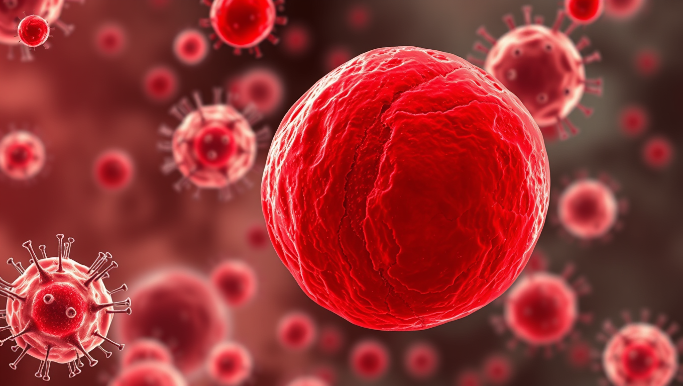
The study has made a groundbreaking discovery about the rapid growth rates of cancerous cells in chronic myeloid leukaemia (CML), a type of blood and bone marrow cancer. Researchers from the Wellcome Sanger Institute used whole genome sequencing to investigate when the abnormal fusion gene BCR::ABL1 arises in a blood cell and how quickly these cells with this genetic change multiply and expand, leading to a diagnosis of CML.

The study, published in Nature, found that the BCR::ABL1 fusion gene typically appears three to 14 years before diagnosis. Once this fusion occurs, the tumour clones grow very quickly, sometimes in excess of 100,000 percent growth annually. This rapid growth is substantially faster than the growth rates of other blood cancers and solid tumours.

Interestingly, age impacts tumour growth rates, with younger patients showing much higher rates at which cancerous cells with the fusion gene multiply compared to older patients. The study also found that patients with faster-growing CML were less likely to respond well to tyrosine kinase inhibitors (TKIs), the standard treatment for CML.

The researchers analyzed sequencing data and health records from over 200,000 participants in the USA-based “All of Us” cohort, showing that almost all individuals with BCR::ABL1 were later diagnosed with a blood disorder. This suggests that expansion of BCR::ABL1 clones without developing subsequent symptoms is unlikely.

Dr Aleksandra Kamizela, co-first author of the study, noted that healthcare professionals can now use this information to better predict patient responses to treatment in a clinical setting. Dr Jyoti Nangalia, senior author of the study, added that CML is an outlier compared to other cancers and that this work paves the way to understanding how to optimise treatment for those patients who currently respond poorly to treatment.

The results have significant implications for the future clinical management of CML, highlighting the importance of considering cancer cell growth rates in a clinical setting.

Alzheimer's

Scientists Uncover Secrets of Protein Blobs That Morph from Liquid to Solid in Human Cells

Scientists examined microscopic blobs of protein found in human cells has discovered that some morph from an almost honey-like substance to a hard candy-like solid. These mysterious droplets, known as biomolecular condensates, solidify when they carry a high proportion of the protein alpha-synuclein, the scientists reported. Clumps of alpha-synuclein are commonly found in the brain cells of people with Parkinson’s disease, a neurodegenerative brain disorder.

Avatar photo

Published

on

The discovery made by an international research collaboration led by Rutgers University-New Brunswick scientists has shed light on the mysteries surrounding biomolecular condensates – microscopic blobs of protein found in human cells. These enigmatic droplets have been observed to morph from a liquid-like substance, similar to honey, into a hard candy-like solid.

Researchers found that these condensates solidify when they contain a high proportion of alpha-synuclein protein, which is commonly associated with the brain cells of individuals suffering from Parkinson’s disease – a neurodegenerative disorder affecting motor control. The study, published in Science Advances, marks a significant breakthrough in understanding the mechanical properties of biomolecular condensates and their link to various biological functions and diseases.

“We can now better comprehend how diseases like Parkinson’s develop and progress by measuring how these condensates change from liquid to solid in living systems,” said Zheng Shi, an assistant professor at the Department of Chemistry and Chemical Biology in the Rutgers School of Arts and Sciences, and senior author of the study. “This knowledge may lead to novel treatments for neurodegenerative diseases.”

The research team employed advanced technologies to achieve a detailed look at biomolecular condensates – structures lacking a membrane boundary. They have designated them as crucial for understanding cell biology and the origins of disease.

Rutgers scientists have successfully developed tools that allow direct, quantitative measurement of material properties in live cells, overcoming previous limitations that only allowed measurements in test tubes. The technique, which takes advantage of the capillary effect, has enabled researchers to pierce condensates with microscopic pipettes (micropipettes) and measure important properties such as viscosity and surface tension.

“This is an exciting technological leap that opens new avenues for research into the early stages of neurodegenerative diseases and their treatment,” Shi said. “Our goal is to continue measuring and better understand the properties of condensates in living cells, which may have significant implications for disease prevention and treatment.”

Other researchers from Rutgers involved in the study included Jean Baum, Mengying Deng, Jordan Elliott, Zhiping Pang, Xiao Su, and Conor McClenaghan.

The discovery made by this research collaboration has sparked new avenues for understanding neurodegenerative diseases and their potential treatments. The findings may ultimately contribute to the development of novel therapeutic strategies that target biomolecular condensates in live cells, offering hope for individuals affected by these debilitating conditions.

Continue Reading

Birth Defects

Breaking Down Barriers: Towards Gene-Targeting Drugs for Brain Diseases

Getting therapeutic drugs past the blood-brain barrier has long been a major challenge in treating brain diseases. Now, researchers have explored how cholesterol-modified heteroduplex oligonucleotides (Chol-HDOs) enhance drug delivery to the brain. Their study reveals that Chol-HDOs bind tightly to serum proteins, allowing them to persist in the bloodstream and cross into brain tissue. These findings offer insights into gene-targeting therapies and could help develop treatments for conditions like Alzheimer’s disease.

Avatar photo

Published

on

Breaking Down Barriers: Towards Gene-Targeting Drugs for Brain Diseases

The human brain is a complex and intricate organ that has long been a challenge to treat when it comes to diseases like Alzheimer’s, Parkinson’s, and brain cancers. One of the major obstacles in delivering therapeutic drugs to the brain is the blood-brain barrier (BBB), a protective layer that restricts the passage of molecules from the bloodstream into the brain.

To overcome this hurdle, researchers at Tokyo University of Science have been exploring new ways to deliver gene-targeting drugs, specifically antisense oligonucleotides (ASOs) and heteroduplex oligonucleotides (HDOs), directly to the brain. In a recent study published in the Journal of Controlled Release, the team led by Professor Makiya Nishikawa demonstrated that modifying HDOs with cholesterol molecules (Chol-HDOs) could improve their stability and specificity, allowing them to penetrate the cerebral cortex beyond the blood vessels.

The key to this success lies in how Chol-HDOs interact with proteins in the bloodstream. Unlike ASOs and HDOs, which bind electrostatically to serum proteins with low affinity and are taken up by cells, Chol-HDOs bind tightly to serum proteins, including lipoproteins, via hydrophobic interactions. This strong binding results in slow clearance from the bloodstream, allowing Chol-HDOs to remain in circulation for a longer period.

The researchers also showed that inhibiting scavenger receptors in cells reduces the uptake of both ASOs and Chol-HDOs in the liver and kidneys, shedding light on how these compounds are taken up by different organs. This finding has significant implications for the design of brain-targeting drugs based on Chol-HDOs.

With over 55 million people living with dementia worldwide and 300,000 cases of brain cancer reported annually, the potential therapeutic applications of modified HDOs are vast. The possibility of efficiently delivering ASOs and other nucleic acid-based drugs to the brain may lead to the development of treatments for brain diseases with significant unmet medical needs.

This study provides valuable insight into how brain-targeting drugs could be designed based on Chol-HDOs, paving the way for a new generation of compounds that effectively target brain diseases. As research continues, we can expect modified HDOs to offer hope to millions of patients and their families around the world.

Continue Reading

Bladder Cancer

Rethinking FOXR2 Activation in Brain Tumors: A Breakthrough in Diagnosis and Care

Scientists have implicated the FOXR2 gene in previously unassociated brain tumor types, with implications for diagnosis, prognosis and treatment.

Avatar photo

Published

on

The world of medicine is abuzz with a groundbreaking discovery that challenges the long-held assumption about the genetic marker FOXR2. For years, physicians have relied on this marker to diagnose central nervous system (CNS) neuroblastoma, a type of brain tumor. However, new research from St. Jude Children’s Research Hospital reveals that FOXR2 activation is not exclusive to CNS neuroblastoma. In fact, it was found in multiple pediatric CNS tumor types, including brain tumors, with significantly different clinical outcomes.

According to Dr. Jason Cheng-Hsuan Chiang, corresponding author of the study, “People have been using FOXR2 activation as a clinical diagnostic for CNS neuroblastoma. But we unexpectedly saw it in a patient’s recurrent non-neuroblastoma tumor, which motivated us to look into other brain tumors.” The researchers used data from the St. Jude Cloud, a vast repository of genomic and sequencing data from St. Jude patients, to identify 42 tumors with activated FOXR2 in 41 patients.

The findings were published today in Neuro-Oncology, a journal of the Society for Neuro-Oncology. “When we looked at the clinical outcomes of the different types of tumors with FOXR2 activation, there was a pretty stark difference,” said co-first author Emily Hanzlik, MD. “The CNS neuroblastomas had an exceptionally good outcome when they were treated with multimodal therapy, whereas the other types of tumors in the cohort, the high-grade gliomas and the pineoblastomas, had pretty dismal outcomes.”

This study highlights the importance of combining molecular findings like DNA and RNA sequencing, histology, and imaging to correctly understand a specific brain tumor. “Only with a holistic view can we choose the best treatment approach for that patient,” Dr. Chiang emphasized.

The discovery of undetected mechanisms of FOXR2 activation in multiple brain tumor types has significant implications for diagnosis, prognosis, and treatment. As Dr. Alexa Siskar, co-first author, noted, “Now that we described these genomic events, hopefully, others will be able to detect them in their patients as well.” This breakthrough opens up new avenues for research and care, ultimately leading to better outcomes for brain tumor patients.

Continue Reading

Trending