Connect with us
We’re experimenting with AI-generated content to help deliver information faster and more efficiently.
While we try to keep things accurate, this content is part of an ongoing experiment and may not always be reliable.
Please double-check important details — we’re not responsible for how the information is used.
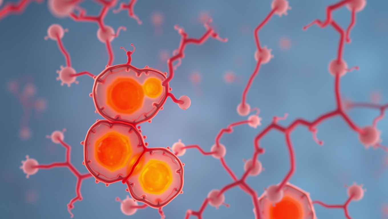
Chronic Illness

Mimicking Embryonic Growth to Break Barriers in Organoid Research

Organoids are made to model human organs and are promising for research and therapy, but there are limitations in their growth and function. A recent study found that placenta-derived IL1 under hypoxic conditions, can greatly increase growth of human stem cell-derived liver organoids. By promoting liver progenitor cell expansion through a specific signaling pathway, this method offers a promising route to improve organoid models and regenerative medicine.

Avatar photo

Published

on

Organoids are small-scale models that mimic human organs. These models have the potential to revolutionize medical research by allowing scientists to understand diseases and test treatments more effectively. Unfortunately, growing organoids that are large and functional enough to be truly useful is a significant challenge. One of the main limitations in organoid growth is the intricate chemical signaling and cellular interactions that occur during fetal development.

Researchers at The University of Tokyo have made an important discovery in this field by finding that placenta-derived IL1α under hypoxic conditions can greatly increase the growth of human stem cell-derived liver organoids. By promoting liver progenitor cell expansion through a specific signaling pathway, this method offers a promising route to improve organoid models and regenerative medicine.

The researchers studied mouse embryo development and found that during a specific stage of liver development (between embryonic days 10 and 11), mouse embryos experience a unique environment characterized by localized blood perfusion and hypoxic conditions. Critically, during this stage, the placenta releases various growth factors that play a pivotal role in the development of the liver.

By identifying and isolating these placental factors, the team focused on a specific protein called IL1α. They introduced this factor to human induced pluripotent stem cell (hiPSC)-derived liver organoids under carefully controlled hypoxic conditions and followed it with controlled oxygenation, mimicking the natural developmental environment. This approach led to remarkable results, as the organoids grew up to five times larger than controls and exhibited improved functional characteristics.

The team demonstrated that placenta-derived IL1α significantly enhanced the proliferation of liver progenitor cells called hepatoblasts. They also performed single-cell RNA sequencing analysis, which revealed that IL1α influences hepatoblast expansion through the SAA1-TLR2-CCL20-CCR6 signaling pathway. These insights provide a clearer understanding of how external factors regulate liver development and offer a novel approach to enhancing organoid growth.

These findings could have significant implications in the medical field. By refining techniques to deliver placenta-derived factors in a controlled manner, future research could pave the way for more advanced organoid-based disease models and potentially facilitate the development of lab-grown organs for transplantation. The team suggests that similar approaches might be applicable to developing organoids for other organ types, opening new frontiers in personalized medicine and regenerative therapies.

While the researchers acknowledge that their approach does not yet fully replicate the dynamic in vivo conditions of fetal liver development, their work marks a critical step toward overcoming existing barriers in organoid research. They suggest that future studies should focus on designing perfusion-based culture systems that can continuously supply placenta-derived factors and oxygen, better simulating the physiological conditions of developing organs.

Overall, by leveraging insights from developmental biology, this research not only enhances our understanding of liver growth but also highlights new pathways for improving the scalability and functionality of hiPSC-derived organoids.

Chronic Illness

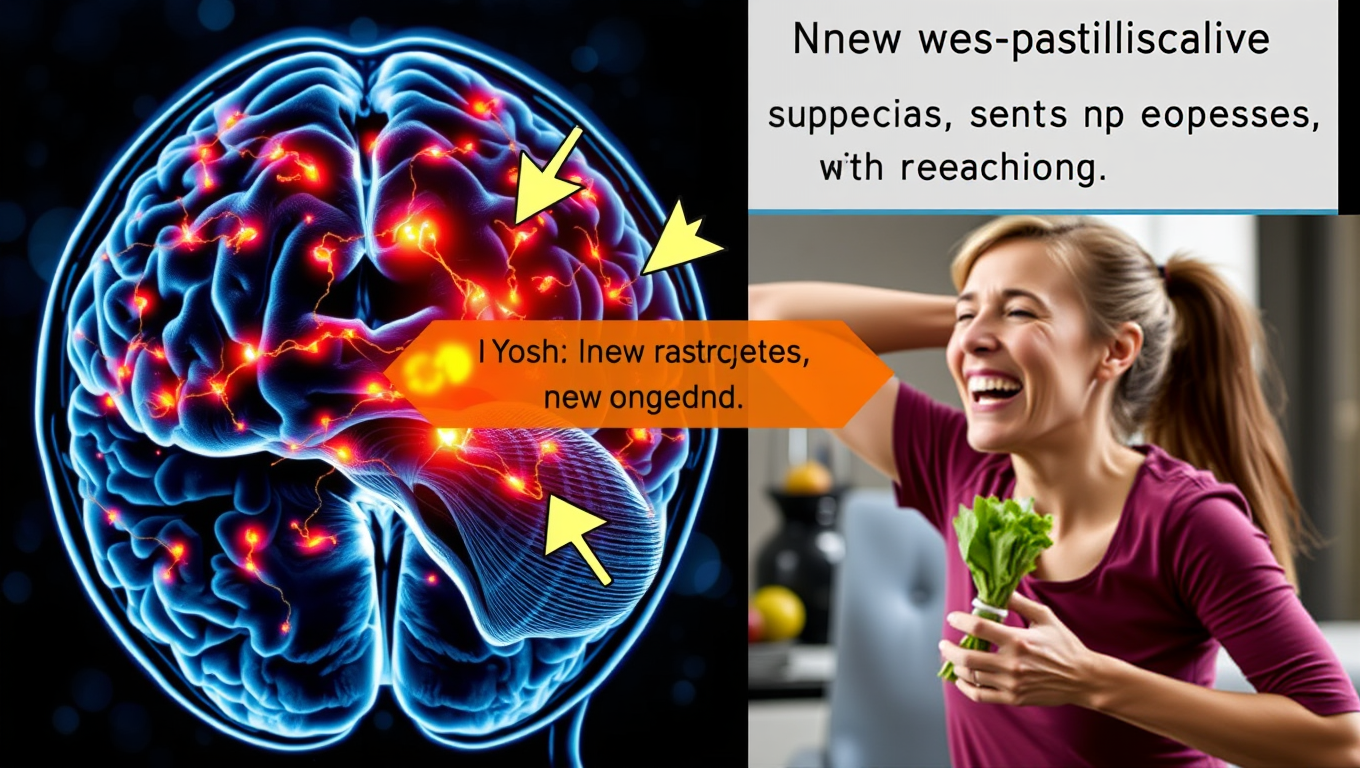
Scientists Uncover Hidden Brain Shortcut for Weight Loss without Nausea

Scientists have uncovered a way to promote weight loss and improve blood sugar control without the unpleasant side effects of current GLP-1 drugs. By shifting focus from neurons to brain support cells that produce appetite-suppressing molecules, they developed a modified compound, TDN, that worked in animal tests without causing nausea or vomiting.
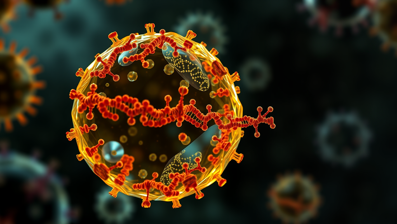
Avatar photo

Published

on

Scientists have made a groundbreaking discovery that could revolutionize the way we approach weight loss. A multidisciplinary team led by Robert Doyle, a chemistry professor at Syracuse University, has identified a hidden brain shortcut that can help people lose weight without experiencing nausea, a common side effect of current weight loss medications.

Current weight loss and diabetes drugs often target brain neurons that control appetite but frequently cause unpleasant side effects like nausea and vomiting. In fact, 70% of patients stop treatment within a year due to these side effects. Doyle’s team has been researching alternative targets for treating obesity and diabetes, looking beyond neurons to study “support” cells such as glia and astrocytes.

The research team discovered that support cells in the hindbrain naturally produce a molecule named octadecaneuropeptide (ODN), which suppresses appetite. In lab tests, injecting ODN directly into rats’ brains made them lose weight and improve how they processed glucose. However, injecting directly into the brain isn’t a practical treatment for people.

To overcome this limitation, researchers created a new version of the molecule named tridecaneuropeptide (TDN), which could be given to human patients through regular injections akin to today’s Ozempic or Zepbound. When tested in obese mice and musk shrews, TDN helped the animals lose weight and respond better to insulin without causing nausea or vomiting.

One goal of the research team is to produce weight loss without aiming new therapeutic molecules at neurons. The new TDN molecule bypasses neurons, taking a shortcut to directly target support cells, which researchers found also produce appetite suppression. This approach has the potential to reduce the unpleasant side effects caused by GLP-1 drugs.

“The idea is to start the process halfway through, reducing the marathon of chemical reactions and negative side effects,” says Doyle. “If we could hit that downstream process directly, then potentially we wouldn’t have to use GLP-1 drugs with their side effects. Or we could reduce their dose, improving the toleration of these drugs.”

A new company called CoronationBio has been launched to turn this discovery into a real-world treatment. The company has licensed intellectual property related to ODN derivatives for the treatment of obesity and cardio-metabolic disease from Syracuse University and the University of Pennsylvania.

Their focus is on translating candidates into the clinic, aiming to start human trials in 2026 or 2027. This breakthrough has the potential to revolutionize the way we approach weight loss, providing a more comfortable and effective solution for millions of people worldwide.

Continue Reading

Cancer

A Silent Killer Unmasked: The Hidden Gene in Leukemia Virus that Could Revolutionize HIV Treatment

Scientists in Japan have discovered a genetic “silencer” within the HTLV-1 virus that helps it stay hidden in the body, evading the immune system for decades. This silencer element essentially turns the virus off, preventing it from triggering symptoms in most carriers. Incredibly, when this silencer was added to HIV, it made that virus less active too — hinting at a revolutionary new strategy for managing not just HTLV-1 but other deadly retroviruses as well. The discovery opens the door to turning the virus’s own stealth tactics against it in future treatments.

Avatar photo

Published

on

A groundbreaking discovery has been made by a research team from Kumamoto University that could revolutionize our understanding and treatment of HIV. The team, led by Professor Yorifumi Satou, has identified a previously unknown genetic “silencer” element in the human T-cell leukemia virus type 1 (HTLV-1) that keeps the virus in a dormant, undetectable state.

HTLV-1 is a cancer-causing retrovirus known to lead to adult T-cell leukemia/lymphoma (ATL), an aggressive and often fatal disease. Despite most infected individuals remaining asymptomatic for life, a fraction eventually develops leukemia or other inflammatory conditions. The virus achieves long-term persistence by entering a “latent” state, where its genetic material hides inside the host’s genome with minimal activity – evading immune detection.

In this study, the research team identified a specific region within the HTLV-1 genome that functions as a viral silencer. This sequence recruits host transcription factors, particularly the RUNX1 complex, which suppresses the virus’s gene expression. When this silencer region was removed or mutated, the virus became more active, leading to greater immune recognition and clearance in lab models.

What’s remarkable is that when the HTLV-1 silencer was artificially inserted into HIV-1 – the virus that causes AIDS – the HIV virus adopted a more latent-like state, with reduced replication and cell killing. This suggests that the silencer mechanism could potentially be harnessed to design better therapies for HIV as well.

“This is the first time we’ve uncovered a built-in mechanism that allows a human leukemia virus to regulate its own invisibility,” said Professor Satou. “It’s a clever evolutionary tactic, and now that we understand it, we might be able to turn the tables in treatment.”

The findings offer hope not only for understanding and treating HTLV-1, especially in endemic regions like southwestern Japan, but also for broader retroviral infections.

Continue Reading

Chronic Illness

The Hidden Link Between Sleep Schedule and Disease Risk

A global study of over 88,000 adults reveals that poor sleep habits—like going to bed inconsistently or having disrupted circadian rhythms—are tied to dramatically higher risks for dozens of diseases, including liver cirrhosis and gangrene. Contrary to common belief, sleeping more than 9 hours wasn’t found to be harmful when measured objectively, exposing flaws in previous research. Scientists now say it’s time to redefine “good sleep” to include regularity, not just duration, as biological mechanisms like inflammation may underlie these powerful sleep-disease links.

Avatar photo

Published

on

The recent publication of a groundbreaking international study has shed new light on the often-overlooked connection between our sleep schedules and the risk of various diseases. The research, led by teams from Peking University and Army Medical University, analyzed objective sleep data from an impressive 88,461 adults in the UK Biobank, revealing significant associations between sleep traits and a staggering 172 diseases.

One of the key findings is that poor sleep regularity – including inconsistent bedtimes and irregular circadian rhythms – is a critical factor in disease risk. The study used actigraphy data to monitor participants’ sleep patterns over an average of 6.8 years, identifying that 92 diseases had more than 20% of their risk attributed to subpar sleep behavior.

Perhaps most concerning are the links between certain sleep habits and specific diseases. For instance, going to bed after 00:30 was found to increase the risk of liver cirrhosis by an alarming 2.57 times, while low interdaily stability (a measure of consistency in daily sleep patterns) raised the risk of gangrene by a staggering 2.61 times.

Interestingly, the study also challenged previous claims that “long sleep” (more than 9 hours) is inherently bad for our health. While subjective reports have suggested links between long sleep and stroke or heart disease, the objective data revealed only one such association – in this case, with an increased risk of certain diseases.

It’s possible that misclassification may be to blame for these previous findings: a shocking 21.67% of participants who reported sleeping more than 9 hours actually slept less than 6 hours, highlighting how often actual sleep time is confused with time spent in bed.

The lead author of the study, Prof. Shengfeng Wang, emphasized that the results underscore the importance of considering sleep regularity beyond just duration. As we strive to maintain good health, it’s essential to prioritize a consistent and predictable sleep schedule – a crucial factor often overlooked until now.

Future research will delve deeper into the causality of these associations and explore the impact of sleep interventions on chronic disease outcomes. By shedding more light on this critical aspect of our overall well-being, we can work towards developing targeted strategies for promoting healthy sleep habits and reducing disease risk.

Continue Reading

Trending