
While we try to keep things accurate, this content is part of an ongoing experiment and may not always be reliable.
Please double-check important details — we’re not responsible for how the information is used.
Child Development
Music Therapy Breakthrough for Brain-Injured Children: A New Tool for Assessing Consciousness
Music could provide a breakthrough in assessing consciousness levels in children who have suffered significant brain injuries, according to new research.


Autism
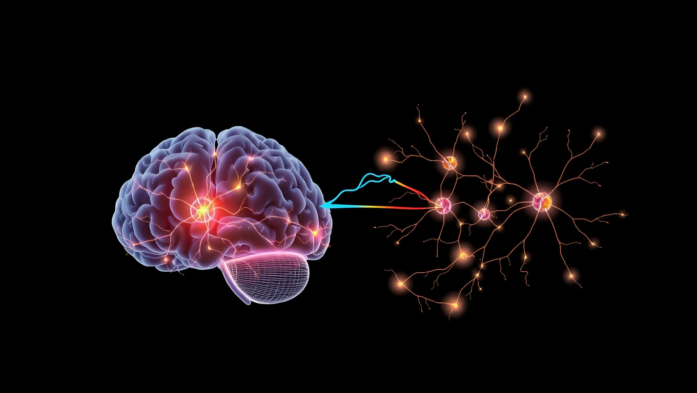
The Thalamic Feedback Loop: Unveiling the Brain’s Secret Pathway to Sensory Perception
Sometimes a gentle touch feels sharp and distinct, other times it fades into the background. This inconsistency isn’t just mood—it’s biology. Scientists found that the thalamus doesn’t just relay sensory signals—it fine-tunes how the brain responds to them, effectively changing what we feel. A hidden receptor in the cortex seems to prime neurons, making them more sensitive to touch.
Child Development
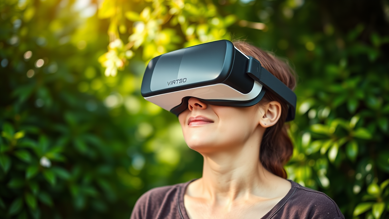
Pain Relief Without Pills? VR Nature Scenes Activate Brain’s Healing Switch
Stepping into a virtual forest or waterfall scene through VR could be the future of pain management. A new study shows that immersive virtual nature dramatically reduces pain sensitivity almost as effectively as medication. Researchers at the University of Exeter found that the more present participants felt in these 360-degree nature experiences, the stronger the pain-relieving effects. Brain scans confirmed that immersive VR scenes activated pain-modulating pathways, revealing that our brains can be coaxed into suppressing pain by simply feeling like we re in nature.
Alternative Medicine
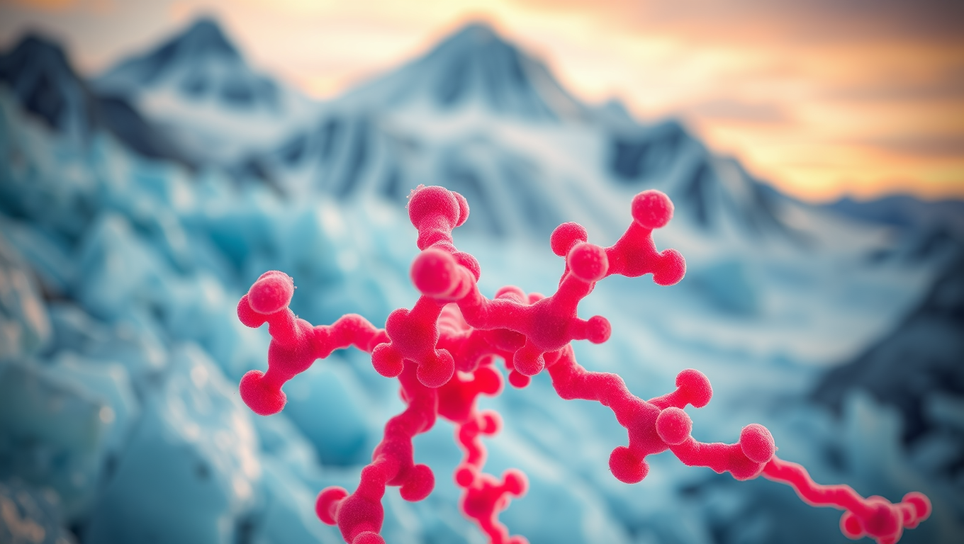
Unlocking the Secrets of Cryorhodopsins: How Arctic Microbes Could Revolutionize Neuroscience
In the frozen reaches of the planet—glaciers, mountaintops, and icy groundwater—scientists have uncovered strange light-sensitive molecules in tiny microbes. These “cryorhodopsins” can respond to light in ways that might let researchers turn brain cells on and off like switches. Some even glow blue, a rare and useful trait for medical applications. These molecules may help the microbes sense dangerous UV light in extreme environments, and scientists believe they could one day power new brain tech, like light-based hearing aids or next-level neuroscience tools—all thanks to proteins that thrive in the cold and shimmer under light.

A New Horizon for Vision: How Gold Nanoparticles May Restore People’s Sight

Retiring Abroad Can Be Lonely Business

Revolutionizing Quantum Communication: Direct Connections Between Multiple Processors
Harnessing Water Waves: A Breakthrough in Controlling Floating Objects

“Unveiling Hidden Patterns: A New Twist on Interference Phenomena”

Household Electricity Three Times More Expensive Than Upcoming ‘Eco-Friendly’ Aviation E-Fuels, Study Reveals
Reducing Falls Among Elderly Women with Polypharmacy through Exercise Intervention
-
 Detectors12 months ago
A New Horizon for Vision: How Gold Nanoparticles May Restore People’s Sight
-
Earth & Climate1 year ago
Retiring Abroad Can Be Lonely Business
-
Cancer1 year ago
Revolutionizing Quantum Communication: Direct Connections Between Multiple Processors
-
Albert Einstein1 year ago
Harnessing Water Waves: A Breakthrough in Controlling Floating Objects
-
Chemistry1 year ago
“Unveiling Hidden Patterns: A New Twist on Interference Phenomena”
-
Earth & Climate1 year ago
Household Electricity Three Times More Expensive Than Upcoming ‘Eco-Friendly’ Aviation E-Fuels, Study Reveals
-
Diseases and Conditions1 year ago
Reducing Falls Among Elderly Women with Polypharmacy through Exercise Intervention
-
Agriculture and Food1 year ago
“A Sustainable Solution: Researchers Create Hybrid Cheese with 25% Pea Protein”


