Connect with us

Brain Injury

The Long-term Effects of Obesity on Brain and Cognitive Health: A Dynamic Relationship Revealed

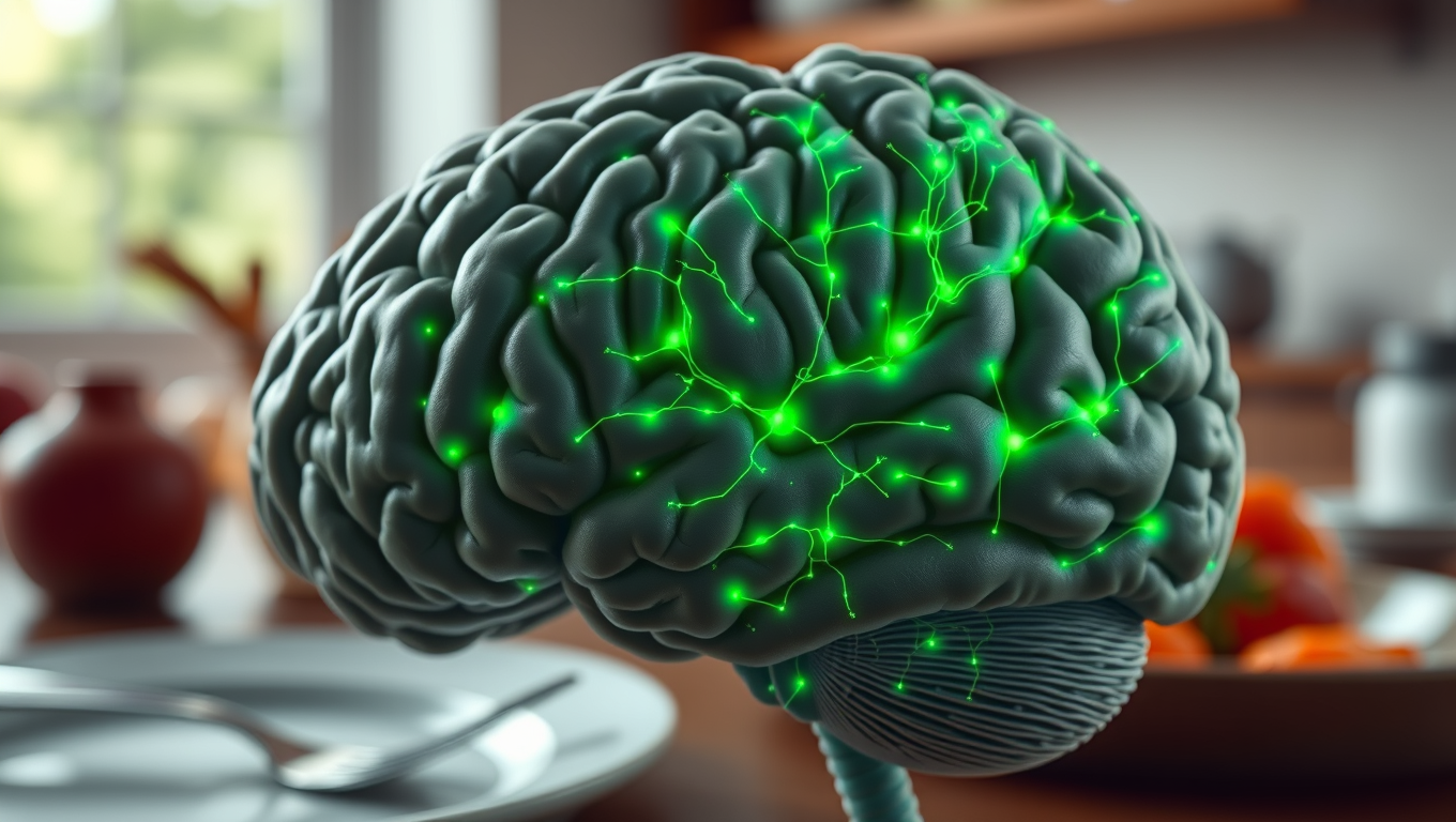
With the global prevalence of obesity on the rise, it is crucial to explore the neural mechanisms linked to obesity and its influence on brain and cognitive health. However, the impact of obesity on the brain is complex and multilevel.
Avatar photo

Published

on

The long-term effects of obesity on brain and cognitive health are a pressing concern, particularly as the global prevalence of obesity continues to rise. A recent study led by Prof. Anqi Qiu has shed new light on this complex relationship, uncovering novel insights into the neural mechanisms underlying the impact of obesity on brain and cognitive aging in adults.

Previous research has primarily relied on cross-sectional samples, making it challenging to distinguish between chronic and temporary obesity. In contrast, Prof. Qiu’s study conducted a comprehensive longitudinal analysis of obesity trajectories and their effects on brain and cognitive health in middle-aged and older adults. This approach allowed researchers to examine how varying levels of obesity over time influence brain anatomy and function differently.

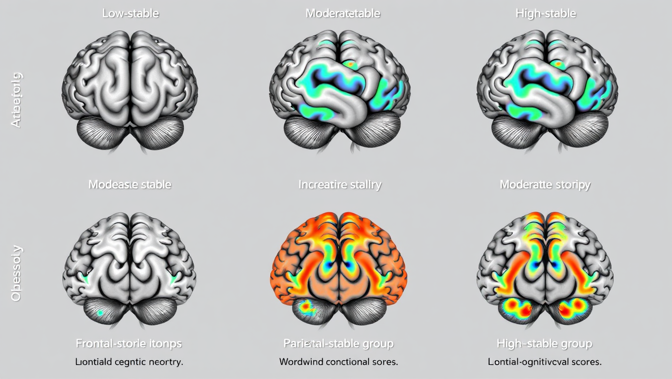
Using multifaceted obesity measurements from the UK Biobank, the study identified five distinct obesity trajectories: low-stable, moderate-stable, high-stable, increasing, and decreasing. Notably, individuals in the decreasing trajectory showed minimal adverse effects on brain structure and cognitive performance compared to those in the low-stable group.

In contrast, the increasing, moderate-stable, and high-stable trajectories were associated with progressively greater impairment in brain structure, functional connectivity, and cognitive abilities. Specifically, these adverse effects began in the fronto-mesolimbic regions for the increasing trajectory, extended to parietal and temporal regions in the moderate-stable group, and ultimately led to widespread brain abnormalities in the high-stable group.

The research highlights the dynamic relationship between obesity progression and brain-cognitive health, underscoring the clinical importance of long-term monitoring and management of obesity through a multifaceted approach. It reveals a distinct, progressive pattern of brain deterioration linked to obesity, suggesting that sustained obesity may accelerate brain aging.

These findings indicate that persistent obesity could serve as a potential biomarker for assessing brain aging. Therefore, reducing both the severity and duration of obesity exposure may be crucial for preserving brain health.

Prof. Qiu stated, “As the aging population grows, there has been a notable rise in neurodegenerative diseases, such as Alzheimer’s disease, Parkinson’s disease, and others, that currently lack a cure. This research proposes that maintaining long-term weight control can contribute to improved brain health.” Looking ahead, the research team plans to integrate multi-omics approaches to explore the biological pathways that influence both brain and body health.

The study, published in Nature Mental Health, extracted obesity measures, brain images, and cognition from the UK Biobank database, which comprised over 500,000 participants aged 40 years and above with diverse ethnic backgrounds.

Brain Injury

Unveiling the Complexity of the Brain’s Serotonin System: A New Frontier in Decision Making

A new study sheds new light on these big questions, illuminating a general principle of neural processing in a mysterious region of the midbrain that is the very origin of our central serotonin (5-HT) system, a key part of the nervous system involved in a remarkable range of cognitive and behavioral functions.

Avatar photo

Published

on

The human brain is a complex organ, comprising billions of neurons that communicate through an intricate network of connections. When it comes to making decisions, our brains engage in a series of binary choices – weighing one option against another. But what happens within our brain when we’re faced with these kinds of decisions? A recent study published in Nature Neuroscience has shed new light on this question, providing compelling insights into the dynamics of the brain’s serotonin system.

Led by researchers at the University of Ottawa Faculty of Medicine, the study reveals that individual serotonin neurons are not independent actors but rather connected to each other through a complex network of axons. This finding challenges the current dominant model, which posits that serotonin neurons operate independently. Instead, the research suggests that distinct groups of serotonin neurons with unique activity patterns control serotonin release in specific regions of the brain.

The study’s first author, Dr. Michael Lynn, emphasizes the significance of this discovery, stating that it could lead to targeted therapeutics for mood disorders like major depressive disorder. The team’s findings also have implications for our understanding of decision-making processes, highlighting a more complex and dynamic set of rules about how and when serotonin is released throughout the brain.

The research has far-reaching consequences for our comprehension of cognitive functions and behavioral outcomes. By identifying a circuit that participates in the computation guiding everyday decisions, the study provides new insights into the neural mechanisms underlying human behavior. As Dr. Jean-Claude Béïque explains, “Do we jump from the high diving board at the pool? Or only from the low one? Do we walk down that very dark alley, or do we avoid it? When is dark too dark?” The answer lies in the intricate computations performed by our brain’s serotonin system.

The research team aims to build on their advances by conducting behavioral studies with mouse models. They hope to replicate the findings in more naturalistic environments, shedding further light on the complex relationships between serotonin release, cognitive functions, and behavioral outcomes. As they continue to explore this new frontier, we can expect a deeper understanding of the brain’s serotonin system and its role in decision-making processes.

Continue Reading

Brain Injury

Favorite Music Triggers a Natural High: Uncovering the Brain’s Secret to Musical Pleasure

A new imaging study showed that listening to favorite music affects the function of the brain’s opioid system.

Avatar photo

Published

on

Favorite music has long been known to evoke intense pleasure, often accompanied by physical sensations such as pleasant “chills.” However, the brain mechanisms behind this phenomenon have remained somewhat of a mystery. Recently, a groundbreaking study conducted at the Turku PET Centre in Finland shed new light on this topic, revealing that listening to favorite music activates the brain’s opioid system.

The study utilized positron emission tomography (PET) and functional magnetic resonance imaging (fMRI) to examine how the brain responds to musical enjoyment. Participants were asked to listen to their favorite music while undergoing these scans, which measured the release of opioids in the brain as well as the density of opioid receptors.

The results showed that listening to favorite music influenced opioid release in several brain areas associated with pleasure, including those linked to the experience of pleasurable chills. Moreover, individual differences in opioid receptor density were found to correlate with brain activation during music listening – the more receptors participants had, the stronger their brains reacted.

According to Academy Research Fellow Vesa Putkinen from the University of Turku, “These results show for the first time directly that listening to music activates the brain’s opioid system. The release of opioids explains why music can produce such strong feelings of pleasure, even though it is not a primary reward necessary for survival or reproduction.”

This study provides significant new insight into how the brain’s chemical systems regulate musical pleasure and may also have practical implications for pain management and mental health treatment. As Professor Lauri Nummenmaa notes, “The brain’s opioid system is involved in pain relief. Based on our findings, the previously observed pain-relieving effects of music may be due to music-induced opioid responses in the brain.”

This rewritten article maintains the core ideas of the original but presents them in a clearer and more engaging manner, making it accessible to a general audience. The prompt for image generation is designed to visually represent the concept of musical pleasure and its neural mechanisms, further emphasizing the key findings of the study.

Continue Reading

Birth Defects

Breaking Down Barriers: Towards Gene-Targeting Drugs for Brain Diseases

Getting therapeutic drugs past the blood-brain barrier has long been a major challenge in treating brain diseases. Now, researchers have explored how cholesterol-modified heteroduplex oligonucleotides (Chol-HDOs) enhance drug delivery to the brain. Their study reveals that Chol-HDOs bind tightly to serum proteins, allowing them to persist in the bloodstream and cross into brain tissue. These findings offer insights into gene-targeting therapies and could help develop treatments for conditions like Alzheimer’s disease.

Avatar photo

Published

on

Breaking Down Barriers: Towards Gene-Targeting Drugs for Brain Diseases

The human brain is a complex and intricate organ that has long been a challenge to treat when it comes to diseases like Alzheimer’s, Parkinson’s, and brain cancers. One of the major obstacles in delivering therapeutic drugs to the brain is the blood-brain barrier (BBB), a protective layer that restricts the passage of molecules from the bloodstream into the brain.

To overcome this hurdle, researchers at Tokyo University of Science have been exploring new ways to deliver gene-targeting drugs, specifically antisense oligonucleotides (ASOs) and heteroduplex oligonucleotides (HDOs), directly to the brain. In a recent study published in the Journal of Controlled Release, the team led by Professor Makiya Nishikawa demonstrated that modifying HDOs with cholesterol molecules (Chol-HDOs) could improve their stability and specificity, allowing them to penetrate the cerebral cortex beyond the blood vessels.

The key to this success lies in how Chol-HDOs interact with proteins in the bloodstream. Unlike ASOs and HDOs, which bind electrostatically to serum proteins with low affinity and are taken up by cells, Chol-HDOs bind tightly to serum proteins, including lipoproteins, via hydrophobic interactions. This strong binding results in slow clearance from the bloodstream, allowing Chol-HDOs to remain in circulation for a longer period.

The researchers also showed that inhibiting scavenger receptors in cells reduces the uptake of both ASOs and Chol-HDOs in the liver and kidneys, shedding light on how these compounds are taken up by different organs. This finding has significant implications for the design of brain-targeting drugs based on Chol-HDOs.

With over 55 million people living with dementia worldwide and 300,000 cases of brain cancer reported annually, the potential therapeutic applications of modified HDOs are vast. The possibility of efficiently delivering ASOs and other nucleic acid-based drugs to the brain may lead to the development of treatments for brain diseases with significant unmet medical needs.

This study provides valuable insight into how brain-targeting drugs could be designed based on Chol-HDOs, paving the way for a new generation of compounds that effectively target brain diseases. As research continues, we can expect modified HDOs to offer hope to millions of patients and their families around the world.

Continue Reading

Trending