Connect with us
We’re experimenting with AI-generated content to help deliver information faster and more efficiently.
While we try to keep things accurate, this content is part of an ongoing experiment and may not always be reliable.
Please double-check important details — we’re not responsible for how the information is used.
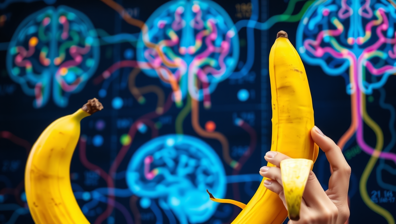
Birth Defects

“The Secret Language of Sight: How Words Shape Our Perception of the World”

Our ability to store information about familiar objects depends on the connection between visual and language processing regions in the brain, according to a new study.

Avatar photo

Published

on

Have you ever stopped to think about how we store information about familiar objects in our brains? A recent study published in PLOS Biology has shed new light on this question, revealing that our ability to recall details like object color is closely linked to the connection between visual and language processing regions in the brain.

The researchers found that when we see an object and know its typical characteristics, such as its usual color, both the visual cortex (which processes what we see) and the language centers of the brain are activated. This connection is crucial for our ability to recall object color knowledge. For example, if you see a yellow banana, your brain’s ventral occipitotemporal cortex (VOTC) is stimulated, not only by the visual information but also by the linguistic association that links the word “banana” to its typical yellow color.

The study used functional magnetic resonance imaging (fMRI) and diffusion imaging to map the white matter connections between language regions and the VOTC in 33 stroke patients and 35 demographically-matched controls. The results showed that stronger connections between visual and language processing regions correlated with better performance on object color knowledge tasks, such as matching objects to their typical colors.

The researchers’ findings have significant implications for our understanding of how we process sensory experiences. They suggest that the connection between vision and language is not just a passive byproduct of brain function but rather an active, dynamic process that shapes how we perceive and store information about the world around us.

As the study’s authors so aptly put it, “Our findings reveal that the brain’s ability to store and retrieve object perceptual knowledge – like the color of a banana – relies on critical connections between visual and language systems. Damage to these connections disrupts both brain activity and behavior, showing that language isn’t just for communication – it fundamentally shapes how sensory experiences are neurally structured into knowledge.”

Autism

The Elusive Science of Tickling: Unraveling the Mysteries of a 2000-Year-Old Enigma

How come you can’t tickle yourself? And why can some people handle tickling perfectly fine while others scream their heads off? Neuroscientists argue that we should take tickle research more seriously.

Avatar photo

Published

on

The science of tickling has been shrouded in mystery for over 2000 years, leaving even the great philosophers Socrates and Charles Darwin baffled. Despite its ubiquity in human interaction, from playful teasing between parents and children to social bonding and emotional expression, the intricacies of tickling remain poorly understood. Neuroscientist Konstantina Kilteni argues that it’s time to take tickle research seriously, shedding light on the complex interplay of motor, social, neurological, developmental, and evolutionary aspects involved.

One of the most intriguing questions surrounding tickling is why we can’t tickle ourselves. Our brain appears to distinguish between self-induced and external stimuli, effectively “switching off” the tickling reflex when we know exactly where and when we’ll be tickled. This phenomenon has sparked interest in understanding what happens in our brain when we’re subjected to ticklish sensations.

Research suggests that people with autism spectrum disorder (ASD) perceive touches as more ticklish than those without ASD, offering a unique window into differences in brain development and function between individuals with and without the condition. Investigating this difference could provide valuable insights into the neurobiology of ASD and potentially inform strategies for better understanding and supporting individuals on the autism spectrum.

From an evolutionary perspective, the purpose and significance of tickling remain unclear. Kilteni notes that even apes like bonobos and gorillas exhibit responses to ticklish touches, while rats have been observed displaying similar behaviors. These observations raise questions about the role of tickling in human evolution and development, as well as its potential functions in social bonding and emotional expression.

To tackle these questions, Kilteni has established a specialized lab dedicated to studying tickling, where researchers can control and replicate various types of ticklish stimuli using mechanical devices like the “tickling chair.” By meticulously recording brain activity and physical reactions such as heart rate, sweating, breathing, laughter, and screaming responses, scientists hope to unlock the secrets of tickling and shed light on its significance in human biology and behavior.

As research continues to unravel the mysteries of tickling, it’s clear that this seemingly simple phenomenon holds a wealth of complexity and intrigue. By taking tickle research seriously, scientists like Kilteni aim to reveal new insights into human brain development, social bonding, emotional expression, and even the intricacies of ASD. The journey ahead promises to be fascinating, as we continue to explore the elusive science of tickling.

Continue Reading

Birth Control

New Method Reduces Risk of Severe Birth Injuries with Accurate Predictions

High birth weight is the main risk factor for birth injuries to the anal sphincter muscles of the person giving birth, according to a new study. This new method for predicting the risks could improve care and reduce injuries.

Avatar photo

Published

on

A recent study conducted at the University of Gothenburg has shed new light on the risk factors for severe birth injuries, specifically obstetric anal sphincter injuries (OASI). The research aimed to develop and validate a prediction model that assesses the risk of OASI before vaginal delivery. This breakthrough could lead to improved care and reduced injuries during childbirth.

In Sweden, five percent of women giving birth to their first child experience OASI, which can have long-term consequences on physical health and quality of life. The study examined registry data from all 45 maternity units in Sweden for the period 2009-2017, involving over 600,000 singleton, head-first births.

The research revealed that larger babies are the strongest predictor of OASI, with a significant increase in risk for those giving birth vaginally for the first time. Previous OASI was also found to be a strong indicator of repeat injury among women having their second vaginal delivery. Furthermore, the use of a vacuum cup during assisted birth was identified as another risk factor.

The prediction models developed by the researchers demonstrated high accuracy and reliability, comparable to established tools used in other fields such as cardiovascular disease and breast cancer. The study’s lead author, Jennie Larsudd-Kåverud, emphasized that these models enable healthcare professionals and pregnant women to assess the risk of severe birth injury together, facilitating joint planning and prevention.

With this new method, medical staff can identify individuals at higher risk of OASI and take necessary precautions to minimize the chances of severe birth injuries. This innovative approach has the potential to improve care during childbirth and reduce long-term health consequences for mothers and their babies.

Continue Reading

Alzheimer's

Unraveling the Mystery of Middle-Age Dementia: Researchers Discover Specific Proteins Linked to Frontotemporal Degeneration (FTD)

Dementia usually affects older people, so when it occurs in middle age, it can be hard to recognize. The most common form is frontotemporal dementia (FTD), which is often mistaken for depression, schizophrenia, or Parkinson’s disease before the correct diagnosis is reached.

Avatar photo

Published

on

In the midst of life, dementia often seems like an affliction that only affects older people. However, for those who suffer from frontotemporal degeneration (FTD), this is not always the case. FTD is a type of dementia that can strike middle-aged individuals, leaving them with symptoms that are often mistaken for depression, schizophrenia, or Parkinson’s disease.

Researchers at the University of California, San Francisco (UCSF) have made a groundbreaking discovery that could lead to better diagnostics and treatment options for patients suffering from FTD. The study, funded by the National Institutes of Health (NIH), analyzed spinal tap fluid from over 4,000 proteins in individuals with inherited forms of FTD.

By comparing these proteins to those found in healthy relatives, the researchers identified specific changes that are associated with FTD. These changes suggest problems with RNA regulation and defects that affect connections in the brain.

“This is a significant breakthrough,” said Rowan Saloner, PhD, professor at UCSF’s Memory and Aging Center. “FTD affects people in the prime of their lives, stripping them of their independence. But there’s no definitive way to diagnose it in living patients, unlike other dementias like Alzheimer’s disease.”

The study’s findings have opened up new possibilities for diagnosing FTD early on and directing patients to relevant therapeutic trials. As Dr. Saloner noted, “If we’re able to identify FTD early on, perhaps using some of the proteins we’ve identified, we can direct patients to the right resources, get them into the right therapeutic trials, and, ultimately, we hope, provide them with precision treatments.”

The research team is led by Kaitlin Casaletto, PhD, professor at UCSF’s Memory and Aging Center, and includes co-authors from the ALLFTD Consortium, which comprises experts from UCSF and the Mayo Clinic. Their study has been published in Nature Aging on May 16.

In conclusion, this groundbreaking research offers hope for patients suffering from FTD. By identifying specific proteins linked to the disease, scientists may be able to develop new diagnostic tools and treatments that can improve the lives of those affected by middle-age dementia.

Continue Reading

Trending