Connect with us
We’re experimenting with AI-generated content to help deliver information faster and more efficiently.
While we try to keep things accurate, this content is part of an ongoing experiment and may not always be reliable.
Please double-check important details — we’re not responsible for how the information is used.

Brain Injury

Uncovering the Silent Thief of Sight: New Study Reveals Link Between Eye Pressure and Vision Loss

One of the world’s leading causes of irreversible vision loss could begin with elevated eye pressure, according to a recent study.
Avatar photo

Published

on

Uncovering the Silent Thief of Sight: New Study Reveals Link Between Eye Pressure and Vision Loss

Glaucoma, a leading cause of irreversible vision loss worldwide, may begin with an often-overlooked factor: elevated eye pressure. A recent study published in the American Academy of Ophthalmology sheds new light on this complex condition, revealing a link between intraocular pressure (IOP) and damage to the optic nerve.

Researchers from the University of Mississippi and the University of Pittsburgh used 3D modeling and fluorescent dye to trace blood flow through the eye under varying amounts of pressure. Their findings suggest that even mildly elevated IOP can distort blood vessels and lead to oxygen deficits, while extreme pressure led to hypoxia in approximately 30% of the lamina cribrosa tissue.

“We wanted to see how intraocular pressure changes and deforms the blood vessels in the eye,” said Yi Hua, a biomedical engineering professor at the University of Mississippi. “If we can understand that, we can inform drug delivery to improve blood flow in the back of the eye. That can slow down the progression of glaucoma.”
Glaucoma damages the optic nerve, leading to irreversible vision loss. It is often referred to as the “silent thief of sight,” with symptoms not becoming apparent until extensive damage has occurred.

The study’s co-author, Yuankai Lu, a postdoctoral researcher at the University of Pittsburgh, noted that previous research had correlated elevated eye pressure with glaucoma but did not explain how those issues were related. By combining imaging techniques with 3D modeling, the researchers gained a more comprehensive understanding of blood flow and oxygen distribution in the eye.
Treatment options are available for elevated eye pressure, but they are most effective for patients who undergo regular eye examinations and are diagnosed early, especially if they are at risk of developing glaucoma, Hua said.

Risk factors include medical conditions like high blood pressure or diabetes, a family history of the disease, and race. Studies show that Black and Latino individuals are more likely to be affected.
“We really want to raise awareness of this issue,” Hua said. “A lot of people know the risk of high blood pressure, but we want to also raise the importance of elevated eye pressure.”
This study highlights the need for better methods to slow down the progression of glaucoma and prevent vision loss. By understanding the relationship between eye pressure and optic nerve damage, researchers can develop new drug pathways and improve treatment options for patients at risk of developing this devastating disease.

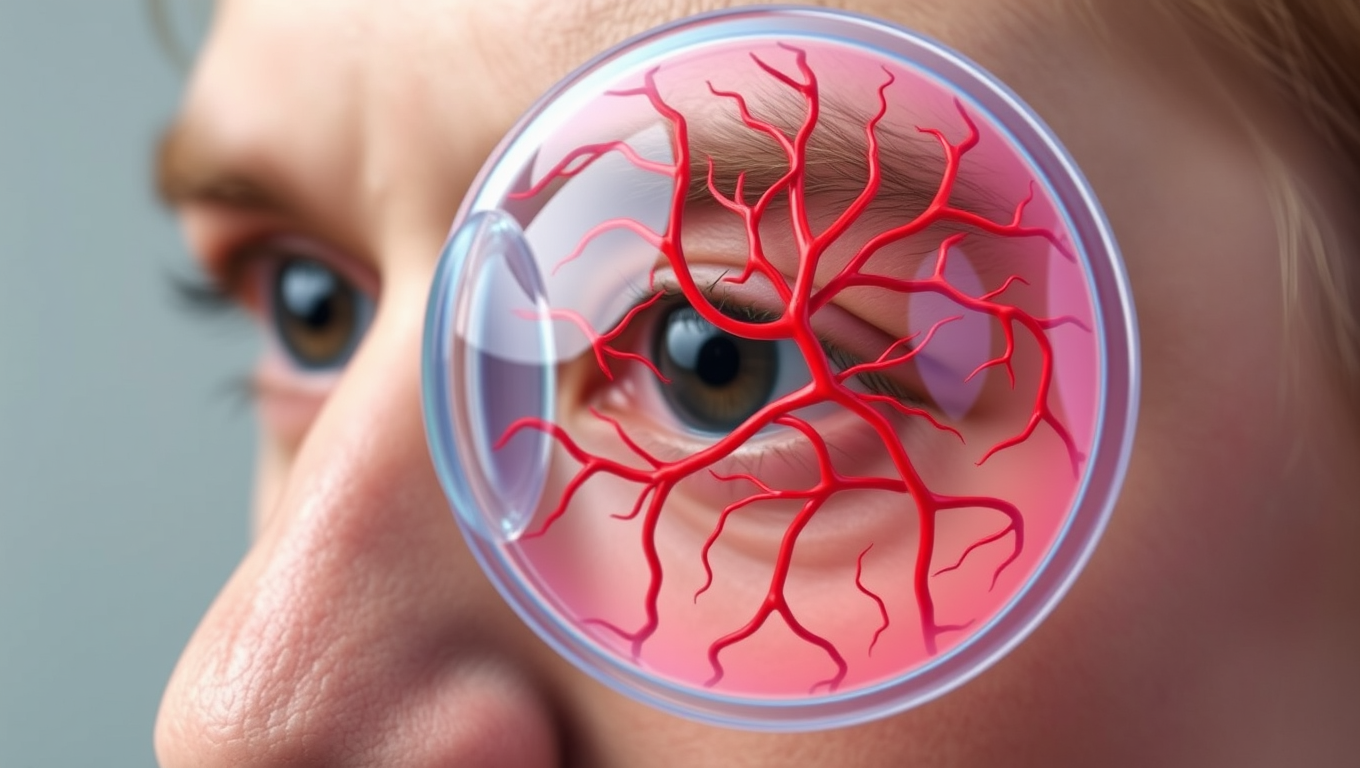
(Note: The image prompt is designed to visually represent the complex relationship between eye pressure and optic nerve damage, highlighting the importance of early diagnosis and treatment.)

Autism

The Brain’s Hidden Patterns: Uncovering the Secret to Flexibility and Stability

A new study challenges a decades-old assumption in neuroscience by showing that the brain uses distinct transmission sites — not a shared site — to achieve different types of plasticity.

Avatar photo

Published

on

By

The Brain’s Hidden Patterns: Uncovering the Secret to Flexibility and Stability

For decades, scientists believed that the brain used a single, shared transmission site for all types of plasticity. However, a groundbreaking study from researchers at the University of Pittsburgh has challenged this assumption, revealing that the brain employs distinct transmission sites to achieve different types of plasticity.

The study, published in Science Advances, offers a deeper understanding of how the brain balances stability with flexibility – a process essential for learning, memory, and mental health. By uncovering the hidden patterns of the brain’s transmission sites, researchers hope to shed light on the underlying mechanisms that govern our thoughts, emotions, and behaviors.

Neurons communicate through synaptic transmission, where one neuron releases chemical messengers called neurotransmitters from a presynaptic terminal. These molecules travel across a microscopic gap called a synaptic cleft and bind to receptors on a neighboring postsynaptic neuron, triggering a response.

Traditionally, scientists believed that spontaneous transmissions (signals that occur randomly) and evoked transmissions (signals triggered by sensory input or experience) originated from one type of canonical synaptic site and relied on shared molecular machinery. However, the research team led by Oliver Schlüter discovered that the brain instead uses separate synaptic transmission sites to carry out regulation of these two types of activity.

The study focused on the primary visual cortex, where cortical visual processing begins. The researchers expected spontaneous and evoked transmissions to follow a similar developmental trajectory, but instead found that they diverged after eye opening.

As the brain began receiving visual input, evoked transmissions continued to strengthen. In contrast, spontaneous transmissions plateaued, suggesting that the brain applies different forms of control to the two signaling modes. To understand why, the researchers applied a chemical that activates otherwise silent receptors on the postsynaptic side, causing spontaneous activity to increase while evoked signals remained unchanged.

This division likely enables the brain to maintain consistent background activity through spontaneous signaling while refining behaviorally relevant pathways through evoked activity. This dual system supports both homeostasis and Hebbian plasticity – the experience-dependent process that strengthens neural connections during learning.

“Our findings reveal a key organizational strategy in the brain,” said Yue Yang, a research associate in the Department of Neuroscience and first author of the study. “By separating these two signaling modes, the brain can remain stable while still being flexible enough to adapt and learn.”

The implications could be broad. Abnormalities in synaptic signaling have been linked to conditions like autism, Alzheimer’s disease, and substance use disorders. A better understanding of how these systems operate in the healthy brain may help researchers identify how they become disrupted in disease.

“Learning how the brain normally separates and regulates different types of signals brings us closer to understanding what might be going wrong in neurological and psychiatric conditions,” said Yang.

Continue Reading

Brain Injury

Brain Training Game Offers New Hope for Drug-Free Pain Management

A trial of an interactive game that trains people to alter their brain waves has shown promise as a treatment for nerve pain — offering hope for a new generation of drug-free treatments.

Avatar photo

Published

on

A groundbreaking trial of an interactive game that trains people to alter their brain waves has shown promise as a treatment for nerve pain – offering hope for a new generation of drug-free treatments.

The PainWaive technology, developed by UNSW Sydney researchers, teaches users how to regulate abnormal brain activity linked to chronic nerve pain, providing a potential in-home, non-invasive alternative to opioids.

A recent trial led by Professor Sylvia Gustin and Dr Negin Hesam-Shariati from UNSW Sydney’s NeuroRecovery Research Hub has delivered promising results, published in the Journal of Pain. The study compared hundreds of measures across participants’ pain and related issues like pain interference before, during, and after four weeks of interactive game play.

Their brain activity was tracked via EEG (electroencephalogram) headsets, with the app responding in real time to shifts in brainwave patterns. Three out of the four participants showed significant reductions in pain, particularly nearing the end of the treatment. Overall, the pain relief achieved by the three was comparable to or greater than that offered by opioids.

“Restrictions in the study’s size, design, and duration limit our ability to generalise the findings or rule out placebo effects,” Dr Hesam-Shariati says. “But the results we’ve seen are exciting and give us confidence to move to the next stage and our larger trial.”

The PainWaive project builds on UNSW Professor Sylvia Gustin’s seminal research into changes in the brain’s thalamus – a central relay hub in the brain – associated with nerve (neuropathic) pain. “The brainwaves of people with neuropathic pain show a distinct pattern: more slow theta waves, fewer alpha waves, and more fast, high beta waves,” Prof. Gustin says.

“We believe these changes interfere with how the thalamus talks to other parts of the brain, especially the sensory motor cortex, which registers pain.” I wondered, can we develop a treatment that directly targets and normalises these abnormal waves?” The challenge was taken up by an interdisciplinary team at UNSW Science and Neuroscience Research Australia (NeuRA), led by Prof. Gustin and Dr Hesam-Shariati, and resulted in PainWaive.

The four participants in its first trial received a kit with a headset and a tablet preloaded with the game app, which includes directions for its use. They were also given tips for different mental strategies, like relaxing or focusing on happy memories, to help bring their brain activity into a more “normal” state. The user data was uploaded to the research team for remote monitoring.

“After just a couple of Zoom sessions, participants were able to run the treatment entirely on their own,” says Dr Hesam-Shariati. “Participants felt empowered to manage their pain in their own environment. That’s a huge part of what makes this special.”

Initially, Dr Hesam-Shariati says, the team planned to use existing commercial EEG systems but were either too expensive or didn’t meet the quality needed to deliver the project. Instead, they developed their own. “Everything except the open-source EEG board was built in-house,” says Dr Hesam-Shariati.

“And soon, even that will be replaced by a custom-designed board.” Thanks to 3D printing, Prof. Gustin says, the team has cut the cost of each headset to around $300 – a fraction of the $1,000 to $20,000 price tags of existing systems.

The headset uses a saline-based wet electrode system to improve signal quality and targets the sensorimotor cortex. “We’ve worked closely with patients to ensure the headset is lightweight, comfortable, and user-friendly,” says Prof. Gustin.

The researchers are now calling for participants to register their interest in two upcoming trials of the neuromodulation technology: the Spinal Pain Trial, investigating its potential to reduce chronic spinal pain, and the StoPain Trial, exploring its use in treating chronic neuropathic pain in people with a spinal cord injury.

Continue Reading

Brain Injury

“Brain Harmony: How Sound Shaping Rewires Your Brain in Real Time”

What happens inside your brain when you hear a steady rhythm or musical tone? According to a new study, your brain doesn’t just hear it — it reorganizes itself in real time.

Avatar photo

Published

on

By

Have you ever wondered what happens inside your brain when you listen to music or a steady rhythm? A groundbreaking study from Aarhus University and the University of Oxford has revealed that your brain doesn’t just hear it – it reorganizes itself in real time. The research, led by Dr. Mattia Rosso and Associate Professor Leonardo Bonetti, introduces a novel neuroimaging method called FREQ-NESS, which maps the brain’s internal organization with high spectral and spatial precision.

Using advanced algorithms, FREQ-NESS disentangles overlapping brain networks based on their dominant frequency, allowing scientists to track how each frequency propagates in space across the brain. This development represents a major advance in neuroscience, enabling researchers to study the brain’s large-scale dynamics more accurately.

The traditional view of brainwaves as fixed stations – alpha, beta, gamma – is being challenged by this research. FREQ-NESS reveals that brain activity is organized through frequency-tuned networks, both internally and externally. This understanding opens new possibilities for basic neuroscience, brain-computer interfaces, and clinical diagnostics.

This study contributes to a growing body of research exploring how the brain’s rhythmic structure shapes perception, attention, and consciousness. Professor Leonardo Bonetti notes, “The brain doesn’t just react – it reconfigures. And now we can see it.” This breakthrough could revolutionize how scientists study brain responses to music and beyond.

A large-scale research program is underway to build on this methodology, supported by an international network of neuroscientists. FREQ-NESS may also pave the way for individualized brain mapping, offering new insights into the intricate harmony of the human brain.

Continue Reading

Trending