Connect with us

Biochemistry

Unlocking the Code: A Breakthrough Tool for Precision Reverse-Engineering of Genetic Programming in Cells

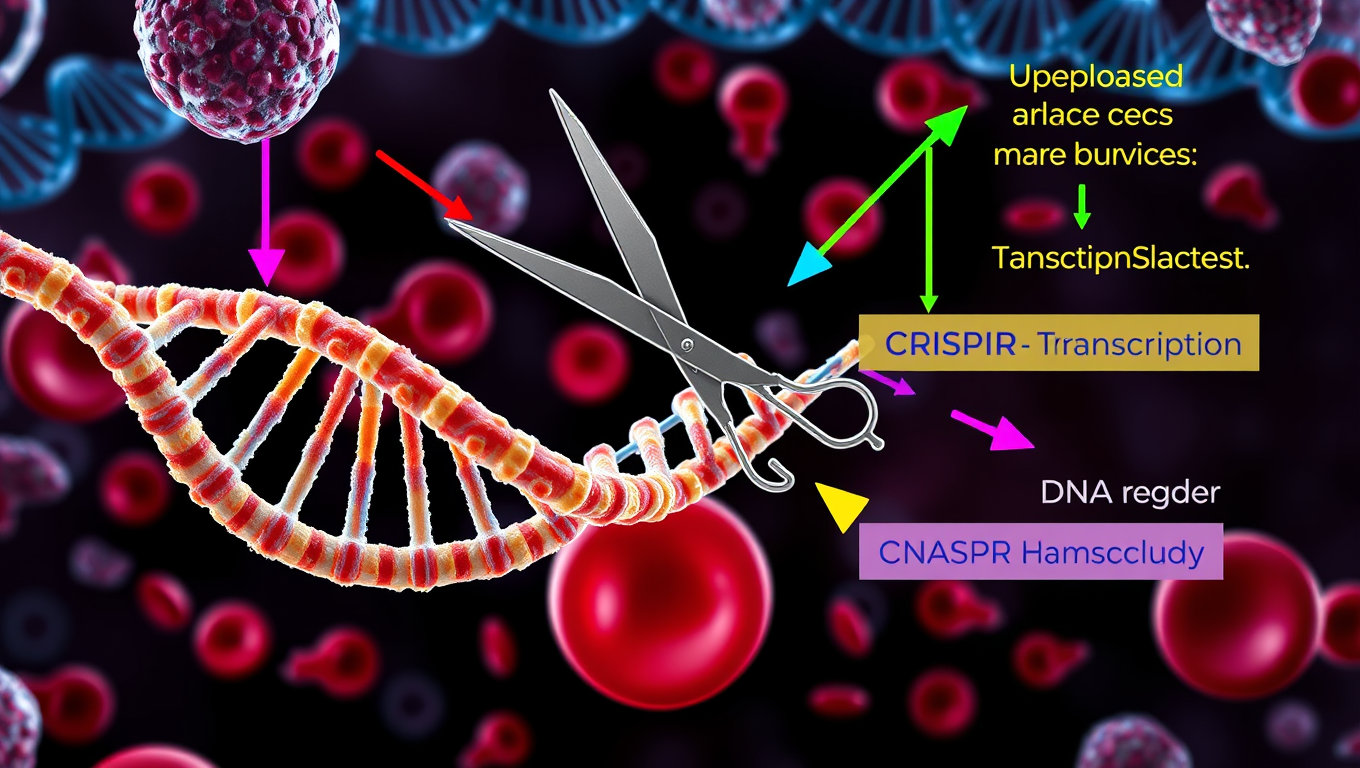
Collaborative research defines a novel approach to understanding how certain proteins called transcription factors determine which genetic programs will drive cell growth and maturation. The method, called ‘Perturb-multiome,’ uses CRISPR to knock out the function of individual transcription factors across many blood cells at once. The researchers then perform single-cell analyses on each cell to measure the effects of the editing, including identifying which genes have been turned on or off and which genes are accessible (based on epigenetic markers).

Avatar photo

Published

on

The scientific community has long sought to understand the intricate mechanisms that govern cellular growth and maturation. A recent collaborative research effort led by investigators at Dana-Farber/Boston Children’s Cancer and Blood Disorders Center has made a significant breakthrough in this regard. The team has developed a novel genomic screening tool called “Perturb-multiome” that enables precision reverse-engineering of genetic programming in cells.

The Perturb-multiome method uses CRISPR technology to knock out the function of individual transcription factors across many blood cells at once. This allows researchers to study the effects of these edits on a single-cell level, including identifying which genes are turned on or off and which ones are accessible based on epigenetic markers. The team applied this tool to immature blood cells to identify the critical transcription factors and DNA regions that strongly influence how blood cells develop.

One of the most significant findings from this study is that many of the DNA regions identified as important for driving blood cell production are also known to harbor mutations linked to blood disorders. These DNA regions occupy less than 0.3% of the genome but explain a disproportionately large share of the genetic influence on blood cell features and specialization.

This new approach has the potential to systematically reveal how thousands of transcription factor variants influence blood cell production and disease risk, creating opportunities for finding many more novel targeted therapies for blood disorders. The implications of this breakthrough are far-reaching, offering hope for patients suffering from various blood-related diseases.

The development of Perturb-multiome is a testament to the power of collaborative research and the importance of investing in scientific inquiry. Funding for this project was provided by several organizations, including La Caixa Foundation, the Rafael del Pino Foundation, the American Society of Hematology, and the National Institutes of Health, among others.

As researchers continue to refine this tool, we can expect even more breakthroughs in our understanding of genetic programming and its role in human health and disease. The future of precision medicine is bright, and the possibilities are endless.

Batteries

“Unlocking the Secrets of Influenza Viruses: How Scientists Are Studying the Interaction between Viruses and Host Cells”

Influenza viruses are among the most likely triggers of future pandemics. A research team has developed a method that can be used to study the interaction of viruses with host cells in unprecedented detail. With the help of their new development, they have also analyzed how novel influenza viruses use alternative receptors to enter target cells.

Avatar photo

Published

on

The threat of future pandemics has been heightened by the emergence of new influenza viruses. In recent years, researchers from the Helmholtz Centre for Infection Research (HZI) and the Medical Center — University of Freiburg have made significant progress in understanding how these viruses interact with host cells.

Led by Professor Christian Sieben’s team at HZI, scientists have developed a novel method to study the initial contact between influenza viruses and host cells. This breakthrough allows researchers to investigate the complex process of viral entry in unprecedented detail.

The researchers immobilized individual viruses on microscopy glass surfaces and then seeded cells on top. This innovative “upside-down” experimental setup enables scientists to analyze the critical moment when viruses interact with cells but do not enter them, stabilizing the initial cell contact for further investigation.

Using high-resolution and super-resolution microscopy, the team demonstrated that contact between the virus and the cell surface triggers a cascade of cellular reactions. The accumulation of local receptors at the binding site, the recruitment of specific proteins, and the dynamic reorganization of the actin cytoskeleton are just some of the processes observed in this study.

What’s more remarkable is that researchers applied their method not only to an established influenza A model but also to a novel strain found in bats. The H18N11 virus, which targets MHC class II complexes rather than glycans on the cell surface, was shown to cluster specific MHCII molecules upon contact with the cell.

This groundbreaking research has significant implications for understanding alternative receptors used by new and emerging influenza viruses. The findings provide a critical basis for investigating potential pandemic pathogens in a more targeted manner, identifying new targets for antiviral therapies, and ultimately developing effective treatments against future pandemics.

The EU project COMBINE, launched in 2025 and coordinated by Professor Sieben’s team at HZI, aims to investigate the virus entry process of newly emerging viruses. This research has far-reaching implications for understanding and combating infectious diseases, making it a significant contribution to the global fight against pandemics.

Continue Reading

Biochemistry

A Breakthrough in Brain Research: The Iontronic Pipette Revolutionizes Neurological Studies

Researchers have developed a new type of pipette that can deliver ions to individual neurons without affecting the sensitive extracellular milieu. Controlling the concentration of different ions can provide important insights into how individual brain cells are affected, and how cells work together. The pipette could also be used for treatments.

Avatar photo

Published

on

The development of an iontronic pipette at Linköping University has opened up new avenues for neurological research. This innovative tool allows researchers to deliver ions directly to individual neurons without affecting the surrounding extracellular milieu. By controlling the concentration of various ions, scientists can gain valuable insights into how brain cells respond to different stimuli and interact with each other.

The human brain consists of approximately 85-100 billion neurons, supported by a similar number of glial cells that provide essential functions such as nutrition, oxygenation, and healing. The extracellular milieu, a fluid-filled space between the cells, plays a crucial role in maintaining cell function. Changes in ion concentration within this environment can activate or inhibit neuronal activity, making it essential to study how local changes affect individual brain cells.

Previous attempts to manipulate the extracellular environment involved pumping liquid into the area, disrupting the delicate biochemical balance and making it difficult to determine whether the substances themselves or the changed pressure were responsible for the observed effects. To overcome this challenge, researchers at the Laboratory of Organic Electronics developed an iontronic micropipette measuring only 2 micrometers in diameter.

This tiny pipette can deliver ions such as potassium and sodium directly into the extracellular milieu, allowing scientists to study how individual neurons respond to these changes. Glial cell activity is also monitored, providing a more comprehensive understanding of brain function.

Theresia Arbring Sjöström, an assistant professor at LOE, highlighted that glial cells are critical components of the brain’s chemical environment and can be precisely activated using this technology. In experiments conducted on mouse hippocampus tissue slices, it was observed that neurons responded dynamically to changes in ion concentration only after glial cell activity had saturated.

This research has significant implications for neurological disease treatment. The iontronic pipette could potentially be used to develop extremely precise treatments for conditions such as epilepsy, where brain function can be disrupted by localized imbalances in ion concentrations.

Researchers are now continuing their studies on chemical signaling in healthy and diseased brain tissue using the iontronic pipette. They also aim to adapt this technology to deliver medical drugs directly to affected areas of the brain, paving the way for more targeted treatments for neurological disorders.

Continue Reading

Biochemistry

Unraveling Nature’s Secret: How Velvet Worm Slime Could Revolutionize Sustainable Materials Design

A new discovery about the slime ejected by velvet worms could revolutionize sustainable material design. The findings outline how a naturally occurring protein structure, conserved across species from Australia, Singapore and Barbados over nearly 400 million years of evolution, enables the slime’s transformation from liquid to fiber and back again. It’s a discovery that could inspire next-generation recyclable bioplastics.

Avatar photo

Published

on

The discovery of the remarkable properties of velvet worm slime has sent shockwaves through the scientific community, offering new hope for sustainable material design. Researchers from McGill University have made a groundbreaking find that could lead to the development of next-generation recyclable bioplastics.

Velvet worms, small caterpillar-like creatures found in humid forests of the southern hemisphere, possess an extraordinary ability – their slime can transform from liquid to fibre and back again. This remarkable property has puzzled scientists for centuries, but a team led by Matthew Harrington, a chemistry professor and Canada Research Chair in green chemistry, has finally decoded the molecular structure behind this phenomenon.

Using protein sequencing and AI-driven structure prediction (AlphaFold), the researchers identified previously unknown proteins in the slime that function similarly to cell receptors in the immune system. These receptor proteins appear to link large structural proteins during fibre formation, enabling the slime’s remarkable reversibility.

The implications of this discovery are profound. Traditional plastics and synthetic fibres require energy-intensive processes to manufacture and recycle, often involving heat or chemical treatments. In contrast, the velvet worm uses simple mechanical forces – pulling and stretching – to generate strong, durable fibres from biorenewable precursors, which can later be dissolved and reused without harmful byproducts.

While a plastic bottle that dissolves in water may seem like an impractical solution, Harrington believes that adjusting the chemistry of this binding mechanism could overcome this limitation. The team’s next challenge will be to experimentally verify the binding interactions and explore whether the principle can be adapted for engineered materials.

The study was co-authored by researchers from McGill University and Nanyang Technological University (NTU) in Singapore, highlighting the importance of international collaboration in addressing pressing global challenges.

As Harrington aptly puts it, “Nature has already figured out a way to make materials that are both strong and recyclable. By decoding the molecular structure of velvet worm slime, we’re now one step closer to replicating that efficiency for the materials we use every day.”

Continue Reading

Trending