Connect with us
We’re experimenting with AI-generated content to help deliver information faster and more efficiently.
While we try to keep things accurate, this content is part of an ongoing experiment and may not always be reliable.
Please double-check important details — we’re not responsible for how the information is used.

Biology

“Unlocking the Secrets of Fish Skin: A Breakthrough in Wound Healing Research”

Scientists have discovered cells in the skin of Atlantic salmon that offer new insights into how wounds heal, tissues regenerate, and cellular transitions support long-term skin health.
Avatar photo

Published

on

The University of Stirling’s Institute of Aquaculture has made a groundbreaking discovery in the field of wound healing research. Scientists have identified a previously unknown population of stem cells in the skin of Atlantic salmon that offers new insights into how wounds heal, tissues regenerate, and cellular transitions support long-term skin health.

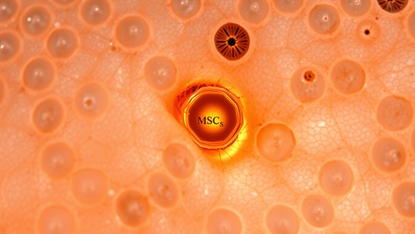
Led by Dr. Rose Ruiz Daniels, the research team used advanced cell profiling technologies to profile skin cells during a wound healing time course. They found fibroblast-like stem cells (MSCs) playing a central role in the remodelling phase of wound healing, which is critical for restoring skin integrity following injury.

The study, published in BMC Biology, reveals that MSCs are not only present at the wound site but also in intact skin, suggesting these adult stem cells are a stable and functional part of salmon skin. They become more transcriptionally active during the remodelling stage of healing and show signs of differentiating into multiple tissue types, including bone and fat.

This discovery has far-reaching implications for aquaculture, as barrier tissue health is a major challenge in Atlantic salmon farming, leading to increased mortality rates in sea cages. By understanding how skin cells remodel and heal tissue, researchers hope to develop new strategies to enhance tissue integrity and reduce non-healing wounds at barrier tissues.

The study is a collaborative effort across institutions, including the University of Stirling, the Roslin Institute, Nofima, and the University of Prince Edward Island. The findings have potentially far-reaching implications for improving fish health and reducing mortality rates in aquaculture.

As Dr. Ruiz Daniels notes, “These findings have potentially far-reaching implications for aquaculture. Barrier tissue health, particularly of the skin and gills, is a major challenge in Atlantic salmon farming and a leading cause of mortality in sea cages.” The research team hopes to develop new strategies to enhance tissue integrity and reduce non-healing wounds at barrier tissues.

This breakthrough in wound healing research has the potential to improve fish health, reduce mortality rates, and promote sustainable aquaculture practices.

Animals

Sugar-Based Sensors Revolutionize Snake Venom Detection

Researchers have published the first example of a synthetic sugar detection test for snake venom, offering a new route to rapid diagnosis and better antivenoms.

Avatar photo

Published

on

The detection of snake venom is a crucial step in treating life-threatening snake bites. According to the World Health Organization (WHO), every five minutes, 50 people are bitten by a snake worldwide, resulting in four permanent disabilities and one death. Traditional methods for diagnosing snake venom rely on antibodies, which have limitations such as high costs, lengthy procedures, and inconsistencies.

Researchers at the University of Warwick have made a groundbreaking discovery that could revolutionize snake venom detection. They have developed a glycopolymer-based ultraviolet-visible (UV-vis) test to detect Western Diamondback Rattlesnake (Crotalus atrox) venom. This new assay is a cheap and rapid alternative to antibody-based approaches, showcasing a version that specifically detects Crotalus atrox venom.

Dr. Alex Baker, lead researcher of the Baker Humanitarian Chemistry Group, explained that snake venoms are complex, making it challenging to detect toxins in the body. However, their research has produced an assay using synthetic sugars that mimic the natural sugar receptors targeted by venom proteins. The team engineered synthetic chains of sugar-like units (glycopolymers) attached to gold nanoparticles to amplify the response and make the reaction visible.

The Western Diamondback Rattlesnake venom binds to specific sugar molecules on red blood cells and platelets, disrupting blood clotting or interfering with immune responses leading to disability and death. The new assay changes color when venom toxins bind to the synthetic sugars, providing a rapid and cheap detection method beyond antibody-based techniques.

Mahdi Hezwani, first author of the research paper, emphasized that this assay could be a game-changer for snake envenomation. The team tested venom from other snake species, such as the Indian Cobra (Naja naja), and found that it did not interact with glycans in the body. This suggests that the new assay may have potential to distinguish between different snake venoms based on their sugar-binding properties.

This is the first example of a diagnosis test using sugars for detecting snake venom in a rapid detection system, building on the work of the Warwick research group using a glyconanoparticle platform in COVID-19 detection. The new assay is faster, cheaper, and easier to store, making it a more practical solution for treating snake bites.

The University of Warwick’s STEM Connect programme has enabled this innovative research, demonstrating the potential for bold and innovative solutions in addressing global health challenges.

Continue Reading

Animal Learning and Intelligence

The Buzz on Caffeine: How it Affects the Sleeping Brain

Coffee can help you stay awake. But what does caffeine actually do to your brain once you’re asleep? Using AI, a team of researchers has an answer: it affects the brain’s ‘criticality’.

Avatar photo

Published

on

The article delves into the intricate relationship between caffeine and the sleeping brain, offering fresh insights from a recent study published in Nature Communications Biology. Researchers from Université de Montréal have shed new light on how caffeine can modify sleep patterns and influence the brain’s recovery during the night.

Led by Philipp Thölke, a research trainee at UdeM’s Cognitive and Computational Neuroscience Laboratory (CoCo Lab), the team used AI and electroencephalography (EEG) to study caffeine’s effects on sleep. Their findings reveal that caffeine increases the complexity of brain signals and enhances brain “criticality” during sleep – a state characterized by balanced order and chaos.

Interestingly, this effect is more pronounced in younger adults, particularly during REM sleep, the phase associated with dreaming. The researchers attribute this finding to a higher density of adenosine receptors in young brains, which naturally decrease with age. Adenosine is a molecule that accumulates throughout the day, causing fatigue.

The study’s lead author, Thölke, notes that caffeine stimulates the brain and pushes it into a state of criticality, where it is more awake, alert, and reactive. However, this state can interfere with rest at night, preventing the brain from relaxing or recovering properly.

The researchers used EEG to record the nocturnal brain activity of 40 healthy adults on two separate nights: one when they consumed caffeine capsules three hours before bedtime and another when they took a placebo at the same time. They applied advanced statistical analysis and artificial intelligence to identify subtle changes in neuronal activity, revealing that caffeine increased the complexity of brain signals during sleep.

The team also discovered striking changes in the brain’s electrical rhythms during sleep: caffeine attenuated slower oscillations such as theta and alpha waves – generally associated with deep, restorative sleep – and stimulated beta wave activity, which is more common during wakefulness and mental engagement.

These findings suggest that even during sleep, the brain remains in a more activated, less restorative state under the influence of caffeine. This change in the brain’s rhythmic activity may help explain why caffeine affects the efficiency with which the brain recovers during the night, with potential consequences for memory processing.

The study’s implications are significant, particularly given the widespread use of caffeine as a daily remedy for fatigue. The researchers stress the importance of understanding its complex effects on brain activity across different age groups and health conditions. They add that further research is needed to clarify how these neural changes affect cognitive health and daily functioning, potentially guiding personalized recommendations for caffeine intake.

Continue Reading

Biology

Unraveling Microtubule Mysteries: Scientists Crack Code on Cellular Scaffolding Secrets

Scientists found out how naturally unstable filaments decide whether to grow or to shorten.

Avatar photo

Published

on

A groundbreaking study has shed new light on the fundamental mechanisms governing microtubule growth within cells. Researchers from Queen Mary University of London and the University of Dundee have made a significant breakthrough by discovering that the ability of tubulin proteins at microtubule ends to connect with each other sideways determines whether a microtubule elongates or shortens.

Microtubules are crucial protein structures that form the internal skeleton of cells, providing structural support and generating dynamic forces that push and pull. These tiny filaments constantly assemble and disassemble by adding or removing tubulin building blocks at their ends. However, the precise rules dictating whether a microtubule grows or shrinks have long remained a mystery due to the complexity and miniature size of their ends.

The collaborative research team has cracked part of this code using advanced computer simulations coupled with innovative imaging techniques. This interdisciplinary approach has allowed them to address this complex biological question from a fresh perspective, bridging physics and biology.

Dr. Vladimir Volkov, co-lead author from Queen Mary University of London, explained the significance of their findings: “Understanding how microtubules grow and shorten is very important – this mechanism underlies division and motility of all our cells. Our results will inform future biomedical research, particularly in areas related to cell growth and cancer.”

Dr. Maxim Igaev, co-lead author from the University of Dundee, highlighted the power of their interdisciplinary approach: “Bridging physics and biology has allowed us to address this complex biological question from a fresh perspective. This synergy not only enriches both fields but also paves the way for discoveries that neither discipline could achieve in isolation.”

This exciting research deepens our understanding of fundamental cellular processes and opens potential new avenues for biomedical research, particularly in areas concerning cell proliferation and the development of treatments for diseases like cancer.

Continue Reading

Trending