Connect with us

Alzheimer's

Unlocking the Secrets of Heart Health through AI-Powered Mammograms

Mammograms, with the help of artificial intelligence (AI) models, may reveal much more than cancer, according to a new study. The findings highlight how these important cancer screening tools can also be used to assess the amount of calcium buildup in the arteries within breast tissue — an indicator of cardiovascular health.
Avatar photo

Published

on

The American College of Cardiology’s Annual Scientific Session (ACC.25) recently showcased a groundbreaking study that sheds new light on the capabilities of mammograms. These essential cancer screening tools have long been used to detect breast cancer, but now, with the aid of artificial intelligence (AI) models, they can also serve as a window into heart health.

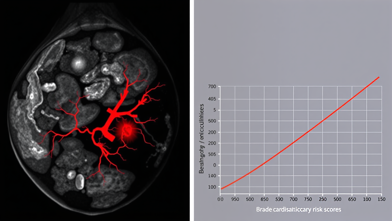
The U.S. Centers for Disease Control and Prevention recommends that middle-aged and older women receive regular mammograms, which are performed over 40 million times annually in the United States. However, radiologists typically do not quantify or report information on breast artery calcifications, which can be seen on these images. This study demonstrates how AI image analysis techniques can automatically analyze breast arterial calcification and provide a cardiovascular risk score.

“We see an opportunity for women to get screened for cancer and also receive a cardiovascular screen from their mammograms,” said Theo Dapamede, MD, PhD, the lead author of the study. The researchers used an AI model to segment calcified vessels in mammogram images and calculate the future risk of cardiovascular events based on data obtained from electronic health record data.

The findings are significant, as heart disease is the leading cause of death in the United States, yet it remains underdiagnosed in women and awareness about this condition lags. By utilizing AI-enabled mammogram screening tools, researchers can identify more women with early signs of cardiovascular disease, taking advantage of routine screenings that many women already receive.

A buildup of calcium in blood vessels is a sign of cardiovascular damage associated with early-stage heart disease or aging. Previous studies have shown that women with calcium buildup in the arteries face a 51% higher risk of heart disease and stroke.

To develop this screening tool, researchers trained a deep-learning AI model on a large dataset, which included images and health records from over 56,000 patients who had a mammogram at Emory Healthcare between 2013 and 2020. The model was then tested for its ability to characterize patients’ cardiovascular risk as low, moderate, or severe based on mammogram images.

The results showed that the AI model performed well in characterizing patients’ cardiovascular risk. After calculating the risk of dying from any cause or suffering an acute heart attack, stroke, or heart failure at two years and five years, the model demonstrated that the rate of these serious cardiovascular events increased with breast arterial calcification level in two of the three age categories assessed – women younger than age 60 and age 60-80, but not in those over age 80.

The researchers also found that women with the highest level of breast arterial calcification (above 40 mm2) had a significantly lower five-year rate of event-free survival compared to those with the lowest level (below 10 mm2). This translates to approximately 2.8 times the risk of death within five years in patients with severe breast arterial calcification compared to those with little to no breast arterial calcification.

The AI model was developed as a collaboration between Emory Healthcare and Mayo Clinic, but it is not currently available for use. If it passes external validation and gains approval from the U.S. Food and Drug Administration, researchers said the tool could be made commercially available for other healthcare systems to incorporate into routine mammogram processing and follow-up care.

The researchers also plan to explore how similar AI models could be used for assessing biomarkers for other conditions, such as peripheral artery disease and kidney disease, that might be extracted from mammograms.

Alzheimer's

Early Menopause Linked to Cognitive Decline: A Study on Women’s Risk Factors

Why does dementia affect more women than men? To help solve this mystery, researchers uncovered a new risk factor: age of menopause onset.

Avatar photo

Published

on

The article “Early Menopause Linked to Cognitive Decline: A Study on Women’s Risk Factors” reveals a significant link between early menopause and cognitive decline in women. Researchers from Tohoku University Graduate School of Medicine and Tokyo Metropolitan Institute of Medical Science conducted a study that analyzed the English Longitudinal Study of Ageing, which included 4,726 women and 4,286 men. The team found that women who entered menopause before the age of 40 had worse cognitive outcomes compared to those who entered menopause after the age of 50.

The researchers were motivated by the disproportionate impact of dementia on women worldwide, as well as the association between early menopause and higher risk of depression in later life. The team controlled for modifiable risk factors for dementia and found that menopause at <40 years was significantly associated with worse cognitive function over a two-year follow-up period. Interestingly, the study also showed that hormone replacement therapy (HRT) did not have an association with cognitive function. This suggests that early menopause may be a direct risk factor for cognitive decline in women. The researchers concluded that understanding this relationship could potentially help design treatments to delay the onset of dementia in at-risk patients. The implications of this study are significant, as it highlights the importance of considering sex-specific factors when assessing the risk of developing dementia. Further research is warranted to elucidate the underlying mechanisms of the relationship between levels of female hormones and cognitive function.

Continue Reading

Alzheimer's

Age, Sex, Hormones and Genetics Uncovered: New Clues on Dementia Biomarkers in the Blood

A new study has found important clues about the roles age, sex, hormonal changes and genetics play in how certain biomarkers for dementia are expressed in the blood, according to a new study.

Avatar photo

Published

on

In a groundbreaking study published in Neurology®, researchers have shed light on how age, sex, hormonal changes, and genetics influence certain biomarkers for dementia in the blood. The study, led by Hannah Stocker, PhD, MPH, of Heidelberg University in Germany, provides valuable insights into the roles these factors play in shaping an individual’s risk of developing dementia.

The researchers analyzed data from a larger 17-year study involving 513 people who developed dementia and 513 who remained free of the condition. The participants had an average age of 64 at the start of the study. By taking blood samples three times during the study, the researchers measured levels of three biomarkers: neurofilament light chain proteins, glial acidic proteins, and phosphorylated tau 181.

The findings revealed that older age was associated with higher levels of all three markers. For example, people aged 75 had an average of 25 picograms per milliliter (pg/ml) for neurofilament light chain proteins compared to those aged 50 who averaged 10 pg/ml. Similarly, glial acidic proteins were found at higher levels in older participants, with a significant difference between those aged 75 and those aged 50.

The study also showed that female participants had higher levels of glial acidic proteins, while male participants had higher levels of neurofilament light chain proteins. Furthermore, the researchers discovered that people who carried the APOEe4 gene had higher levels of tau and glial acidic proteins.

Notably, the study found that female participants who had not yet gone through menopause had higher levels of glial acidic proteins. This may be attributed to having higher levels of sex hormones, which have been linked to neuroinflammation in previous studies.

The findings of this study highlight the importance of further exploring these biomarkers, including during menopause, in the development of dementia. By gaining a better understanding of how age, sex, hormonal changes, and genetics interact with biomarker levels, researchers can improve their ability to test for dementia using simple blood tests.

Continue Reading

Alzheimer's

Breaking Down Brain Inflammation: Scientists Identify Key Enzyme in Alzheimer’s Disease

A research team has identified a previously unknown enzyme, SIRT2, that plays a key role in memory loss associated with Alzheimer’s disease (AD). The study provides critical insights into how astrocytes contribute to cognitive decline by producing excessive amounts of the inhibitory neurotransmitter GABA.

Avatar photo

Published

on

As researchers continue to unravel the mysteries of Alzheimer’s disease (AD), a team at the Institute for Basic Science (IBS) has made a significant breakthrough. They have identified a previously unknown enzyme, SIRT2, that plays a crucial role in memory loss associated with AD. This discovery provides critical insights into how astrocytes contribute to cognitive decline by producing excessive amounts of the inhibitory neurotransmitter GABA.

Astrocytes, once thought to only support neurons, are now known to actively influence brain function. In AD, astrocytes become reactive, attempting to clear amyloid-beta (Aβ) plaques, a hallmark of the disease. However, this process triggers a harmful chain reaction, leading to the overproduction of GABA, which dampens brain activity and causes memory impairment.

The IBS research team used molecular analysis, microscopic imaging, and electrophysiology to identify SIRT2 as one of the critical enzymes involved in GABA overproduction in AD-affected astrocytes. They found that SIRT2 protein was increased in the astrocytes of a commonly used AD mouse model and in post-mortem human AD patient brains.

“When we inhibited the astrocytic expression of SIRT2 in AD mice, we observed partial recovery of memory and reduced GABA production,” said Mridula Bhalla, the lead author of the study. “While we expected reduced GABA release, we found that only short-term working memory was recovered, and spatial memory was not. This was exciting but also left us with more questions.”
The research team’s findings suggest that SIRT2 participates in the last step of GABA production, while hydrogen peroxide (H2O2) is produced earlier in the process. They noted that inhibition of SIRT2 continued H2O2 production, indicating that neuronal degeneration might continue even though GABA production is reduced.

By identifying SIRT2 and ALDH1A1 as downstream targets, scientists can now selectively inhibit GABA production without affecting H2O2 levels. This breakthrough allows researchers to separate the effects of GABA and H2O2 and study their individual roles in neurodegeneration.

“This finding paves the way for more precise therapeutic strategies aimed at controlling astrocytic reactivity in Alzheimer’s disease,” said Director C Justin Lee. “We can now dissect the effects of GABA and H2O2 and study their individual roles in disease progression.” While SIRT2 may not be a direct drug target due to its limited effects on neurodegeneration, this research opens up new possibilities for treating AD.

Continue Reading

Trending