Connect with us
We’re experimenting with AI-generated content to help deliver information faster and more efficiently.
While we try to keep things accurate, this content is part of an ongoing experiment and may not always be reliable.
Please double-check important details — we’re not responsible for how the information is used.

Chronic Illness

Unraveling Memory Formation: A Computational Model Reveals New Insights into Protein Structures at Synapses

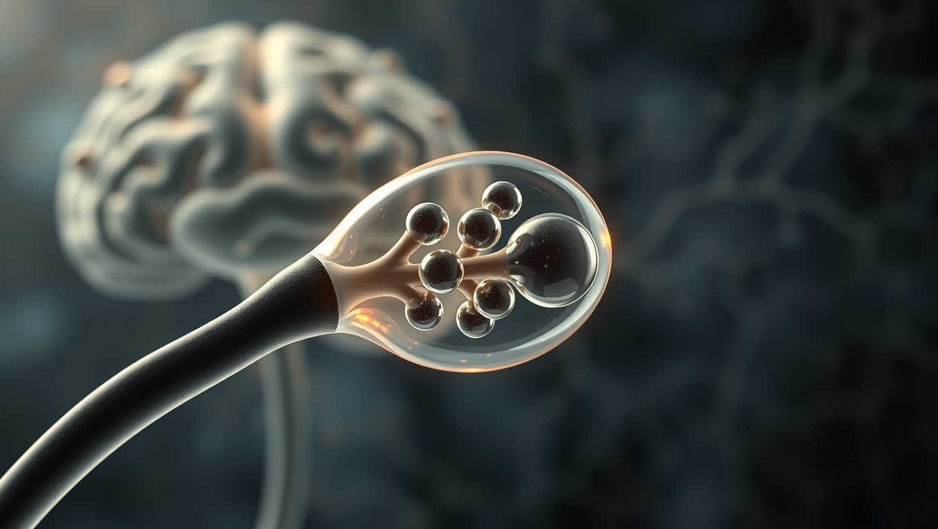
Complex protein interactions at synapses are essential for memory formation in our brains, but the mechanisms behind these processes remain poorly understood. Now, researchers have developed a computational model revealing new insights into the unique droplet-inside-droplet structures that memory-related proteins form at synapses. They discovered that the shape characteristics of a memory-related protein are crucial for the formation of these structures, which could shed light on the nature of various neurological disorders.
Avatar photo

Published

on

Memory formation is one of the brain’s most fundamental and complex functions, yet the microscopic mechanisms behind it remain poorly understood. Recent research has highlighted the importance of biochemical reactions occurring at postsynaptic densities – specialized areas where neurons connect and communicate. These tiny junctions between brain cells are now thought to be crucial sites where proteins need to organize in specific ways to facilitate learning and memory formation.

A 2021 study revealed that memory-related proteins can bind together to form droplet-like structures at postsynaptic densities, which scientists believe may be fundamental to how our brains create lasting memories. However, understanding exactly how and why such complex protein arrangements form has remained a significant challenge in neuroscience.

Against this backdrop, a research team led by Researcher Vikas Pandey from the International Center for Brain Science (ICBS), Fujita Health University, Japan, has developed an innovative computational model that reproduces these intricate protein structures. Their paper, published online in Cell Reports on April 07, 2025, explores the mechanisms behind the formation of multilayered protein condensates.

The researchers focused on four proteins found at synapses, with special attention to Ca²⁺/calmodulin-dependent protein kinase II (CaMKII) – a protein particularly abundant in postsynaptic densities. Using computational modeling techniques, they simulated how these proteins interact and organize themselves under various conditions. Their model successfully reproduced the formation of the above-mentioned “droplet-inside-droplet” structures observed in earlier experiments.

Through simulations and detailed analyses of the physical forces and chemical interactions involved, the research team shed light on a process called liquid-liquid phase separation (LLPS); it involves proteins spontaneously organizing into condensates without membranes that sometimes resemble the organelles found inside cells. Crucially, the researchers found that the distinctive “droplet-inside-droplet” structure appears as a result of competitive binding between the proteins and is significantly influenced by the shape of CaMKII, specifically its high valency (number of binding sites) and short linker length.

These findings could pave the way toward a better understanding of the possible mechanisms of memory formation in humans. However, the long-term implications of this research extend well beyond basic neuroscience. Defects in synapse formation have been associated with numerous neurological and mental health conditions, including schizophrenia, autism spectrum disorders, Down syndrome, and Rett syndrome.

“Our results revealed new structure-function relationships between proteins at synapses,” said Dr. Pandey. “We hope that our findings will contribute to the development of novel therapeutic strategies for these devastating diseases.”

The project received funding from various organizations, including the Core Research for Evolutional Science and Technology (CREST), the Japan Science and Technology Agency (JST), JSPS KAKENHI, Kobayashi foundation, ISHIZUE2024 of Kyoto University, Grant-in-Aid for Scientific Research JP18H05434, and others.

References:

* Pandey, V., et al. (2025). Unraveling memory formation: A computational model reveals new insights into protein structures at synapses. Cell Reports.
* Japanese Ministry of Education, Culture, Sports, Science, and Technology (MEXT). (n.d.). Research Grants JP18H05434 and JP20K21462.

Chronic Illness

Unraveling the Mystery of Stress Granules in Neurodegenerative Diseases

Scientists found that stabilizing stress granules suppresses the effects of ALS-causing mutations, correcting previous models that imply stress granules promote amyloid formation.

Avatar photo

Published

on

The study, led by researchers from St. Jude Children’s Research Hospital and Washington University in St. Louis, has made significant strides in understanding the role of biomolecular condensation in the development of neurodegenerative diseases. The research focuses on the interactions that drive the formation of condensates versus amyloid fibrils and their relationship to stress granules.

Stress granules are temporary structures formed by cells under conditions of cellular stress, akin to a ship lowering its sails in a storm. They have been previously implicated as drivers of neurodegenerative diseases such as Amyotrophic Lateral Sclerosis (ALS) and Frontotemporal Dementia (FTD). The researchers demonstrated that fibrils are the globally stable states of driver proteins, whereas condensates are metastable sinks.

Their findings show that disease-linked mutations diminish condensate metastability, thereby enhancing fibril formation. This suggests that stress granules may not be the primary culprits behind neurodegenerative diseases but rather a protective barrier against them. The researchers also discovered that while fibrils can form on condensates’ surfaces, proteins eventually incorporated into these fibrils stem from outside the condensate.

These discoveries have significant implications for developing potential treatments against neurodegenerative diseases. As lead researcher Tanja Mittag noted, “This information will aid in deciding how to develop potential treatments against a whole spectrum of neurodegenerative diseases.” The study’s findings also highlight the importance of considering stress granules as a protective barrier rather than a crucible for fibril formation.

In conclusion, this research provides crucial insights into the role of stress granules in neurodegenerative diseases. By understanding how these structures interact with fibrils and their relationship to disease-causing mutations, scientists can develop novel therapeutic approaches that may help combat these devastating conditions.

Continue Reading

Child Development

Unpacking the Pace of Aging: A New Tool for Understanding Population Healthspan and Lifespan

A newly refined method for measuring the Pace of Aging in population-based studies provides a powerful tool for predicting risks associated with aging, including chronic illness, cognitive impairment, disability, and mortality. The method offers researchers and policy makers a novel approach to quantify how quickly individuals and populations experience age-related health decline.

Avatar photo

Published

on

The Pace of Aging, a newly refined method for measuring the rate at which individuals experience age-related health decline, offers researchers and policymakers a powerful tool for predicting risks associated with aging, including chronic illness, cognitive impairment, disability, and mortality. Developed by Columbia University Mailman School of Public Health researchers, this approach quantifies how quickly individuals and populations experience age-related health decline.

The existing toolkit for population health research on aging did not distinguish differences caused by early-life factors from those caused by ongoing changes in people’s bodies due to aging. The findings from the study published in Nature Aging highlight the importance of this new method.

“Our Pace of Aging method is an essential approach for understanding population aging,” explained Arun Balachandran, PhD, a postdoctoral researcher at the Columbia Aging Center and lead author of the study. “Our existing toolkit doesn’t include methods that can separate out the legacies of early life from the changes caused by aging.”

Daniel Belsky, PhD, associate professor of Epidemiology at Columbia Mailman School and member of the Robert N. Butler Columbia Aging Center, elaborated on this point: “We developed the Pace of Aging method to evaluate the effectiveness of interventions targeting the biology of aging. The new approach introduced in this paper is designed to do the same for social policies and public health programs.”

The researchers analyzed data from two large-scale studies: the U.S. Health and Retirement Study (HRS) and the English Longitudinal Study of Aging (ELSA). These long-term studies follow adults aged 50 and older, along with their spouses, collecting detailed information on health, cognition, socioeconomic status, and family dynamics.

The new approach makes use of data from dried blood spots, physical exams, and performance tests given to participants in their homes at up to three timepoints over eight-year follow-up intervals. The researchers examined the Pace of Aging in 19,045 participants who contributed data over 2006-2016, with additional follow-up to determine disease, disability, and mortality through 2022.

In the US study, the Pace of Aging was measured from C-reactive protein (CRP), Cystatin-C, glycated hemoglobin (HbA1C), diastolic blood pressure, waist circumference, lung capacity (peak flow), balance, grip strength, and gait speed. The researchers found that their method can measure important variability in the pace of aging in older people with a relatively limited set of measurements.

“Our findings establish that we can measure important variability in the pace of aging in older people with a relatively limited set of measurements,” said Belsky. “Our findings open up possibilities to study pace of aging in cohorts around the world.”

The researchers also found signs of accelerated aging in people with lower levels of education and reported differences in aging trajectories across population subgroups. Originally developed using data from the Dunedin Study, a longitudinal study of individuals born in 1972-73, the initial Pace of Aging tool focused on changes from young adulthood through midlife.

The newly adapted method extends its utility to population-based studies of aging, offering planners and policymakers a valuable resource for monitoring and improving population health and longevity. “Beyond medicine and gerontology, this work has important implications for sociology and economics,” added Belsky.

“It can help us understand how life transitions – such as retirement, caregiving, and bereavement – affect the aging process and support the development of more effective public health and social policies.” The researchers highlighted that the differences in aging speed found were not just statistically significant but meaningful, with people aging faster much more likely to get sick, become disabled, or die sooner.

The study was supported by National Institutes of Health grants R01AG061378, T32AI114398, and U01AG009740; the Russel Sage Foundation; BioSS Grant 1810-08987; and the Robert N. Butler Columbia Aging Center.

Continue Reading

Children's Health

Uncovering Early Signs of Teen Depression through Blood Markers

Using a novel lab method they developed, researchers have identified nine molecules in the blood that were elevated in teens diagnosed with depression. These molecules also predicted how symptoms might progress over time. The findings of the clinical study could pave the way for earlier detection, before symptoms worsen and become hard to treat.

Avatar photo

Published

on

The discovery of nine specific molecules in the blood has revolutionized our understanding of teen depression. McGill University researchers have developed a novel lab method to detect these molecules, known as microRNAs, which can predict how symptoms might progress over time. This breakthrough could lead to earlier detection and intervention before symptoms worsen and become harder to treat.

The alarming rise in adolescent depression diagnoses has severe consequences, including long-lasting effects on mental health, substance use, social isolation, and treatment resistance. By identifying unique microRNA biomarkers linked specifically to teens, researchers hope to provide an additional objective metric for early identification and care.

A minimally invasive approach was used to collect small blood samples from 62 teenagers, 34 with depression and 28 without. The McGill team developed the lab method to extract and analyze microRNAs from these samples, making it practical and scalable for broader use.

The study’s findings pave the way for using dried blood spots as a tool in psychiatric research, allowing us to track early biological changes linked to mental health using a minimally invasive method. Researchers plan to validate their findings in larger groups of adolescents and explore how these microRNAs interact with genetic and environmental risk factors.

The study was funded by various organizations, including the Douglas Foundation, the National Institute on Drug Abuse, and the Canadian Institutes of Health Research.

Continue Reading

Trending