Connect with us
We’re experimenting with AI-generated content to help deliver information faster and more efficiently.
While we try to keep things accurate, this content is part of an ongoing experiment and may not always be reliable.
Please double-check important details — we’re not responsible for how the information is used.

Alzheimer's

Breaking Ground in ALS Research: Uncovering Early Signs of Disease and New Treatment Targets

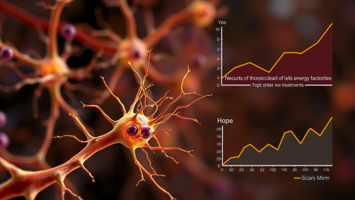
Using the gene scissors CRISPR and stem cells, researchers have managed to identify a common denominator for different gene mutations that all cause the neurological disease ALS. The research shows that ALS-linked dysfunction occurs in the energy factories of nerve cells, the mitochondria, before the cells show other signs of disease, which was not previously known.
Avatar photo

Published

on

Breaking Ground in ALS Research: Uncovering Early Signs of Disease and New Treatment Targets

Researchers at Stockholm University and the UK Dementia Research Institute (UK DRI) have made a groundbreaking discovery that could revolutionize the treatment of Amyotrophic Lateral Sclerosis (ALS). The study, published in Nature Communications, reveals that ALS-linked dysfunction occurs in the energy factories of nerve cells, called mitochondria, before the cells show other signs of disease.

“We show that the nerve cells, termed motor neurons, that will eventually die in ALS have problems soon after they are formed. We saw the earliest sign of problems in the cell’s energy factories, the mitochondria, and also in how they are transported out into the nerve cells’ long processes where there is a great need for them and the energy they produce,” says Dr Eva Hedlund at Stockholm University, head of the study together with Dr Marc-David Ruepp at the UK Dementia Research Institute.

The research team used the gene scissors CRISPR/Cas9 to introduce various ALS-causing mutations into human stem cells, called iPS cells. From these, motor neurons and interneurons were produced, which were then analyzed with single-cell RNA sequencing. The data obtained showed a common disease signature across all ALS-causing mutations, unique to motor neurons.

This happened very early and was completely independent of whether the disease-causing mutated proteins (FUS, or TDP-43) were in the wrong place in the cell or not. “Until now, it has been believed that it is the change where the proteins are within the cells, called mislocalization, that occurs first,” says Dr Marc-David Ruepp.

The researchers also found that most errors arising from ALS-causing mutations are caused by a new toxic property of the protein, not by a loss of function. A third discovery was that the transport of mitochondria out into the axons is radically affected in the ALS lines, independently of whether the disease-causing proteins were in the wrong place in the cell or not.

The new discoveries open up for early treatment methods, something that for the research team is a continuous work in progress. “We are trying to understand how these early errors occur in the sensitive motor neurons in ALS, and how it affects energy levels in the cells and their communication and necessary contacts with muscle fibers,” says Dr Eva Hedlund.

The study’s findings have significant implications for ALS treatment and provide new targets for therapies. The research team is now working on understanding the mechanisms behind these early errors and how to address them, hoping to improve the lives of people affected by this devastating disease.

Alzheimer's

“Unlocking Brain Health: Scientists Discover Key Receptor for Microglia to Fight Alzheimer’s”

Scientists at UCSF have uncovered how certain immune cells in the brain, called microglia, can effectively digest toxic amyloid beta plaques that cause Alzheimer’s. They identified a key receptor, ADGRG1, that enables this protective action. When microglia lack this receptor, plaque builds up quickly, causing memory loss and brain damage. But when the receptor is present, it seems to help keep Alzheimer’s symptoms mild. Since ADGRG1 belongs to a drug-friendly family of receptors, this opens the door to future therapies that could enhance brain immunity and protect against Alzheimer’s in more people.

Avatar photo

Published

on

The scientific community has made significant strides in understanding the complex mechanisms behind Alzheimer’s disease. A recent study by researchers at UC San Francisco has shed light on how microglia, immune cells that play a crucial role in maintaining brain health, can break down and remove toxic proteins associated with the disease. This groundbreaking discovery could pave the way for novel therapeutic approaches to combat Alzheimer’s.

In Alzheimer’s, proteins like amyloid beta clump together, forming plaques that damage the brain. However, in some individuals, microglia effectively engulf and digest these proteins before they can cause harm. The resulting few and smaller clumps are associated with milder symptoms. Researchers at UCSF identified a molecular receptor, ADGRG1, which enables microglia to perform this critical function.

Using a mouse model of Alzheimer’s disease, the researchers observed that the loss of ADGRG1 led to a rapid buildup of amyloid plaques, neurodegeneration, and problems with learning and memory. The study also reanalyzed data from a prior human brain expression study, finding that individuals who died of mild Alzheimer’s had microglia with abundant ADGRG1, whereas those with severe Alzheimer’s had very little ADGRG1.

This discovery has significant implications for the development of new therapies. Since ADGRG1 is one of hundreds of G protein-coupled receptors targeted in drug development, it may be feasible to rapidly translate this finding into new treatments. As Dr. Piao noted, “Some people are lucky to have responsible microglia, but this discovery creates an opportunity to develop drugs to make microglia effective against amyloid-beta in everyone.” The potential for breakthrough therapies is exciting news for those affected by Alzheimer’s and their loved ones.

Continue Reading

Alzheimer's

Rewinding Stroke Damage and Beyond: The Promise of GAI-17

Stroke kills millions, but Osaka researchers have unveiled GAI-17, a drug that halts toxic GAPDH clumping, slashes brain damage and paralysis in mice—even when given six hours post-stroke—and shows no major side effects, hinting at a single therapy that could also tackle Alzheimer’s and other tough neurological disorders.

Avatar photo

Published

on

The devastating effects of stroke can be irreversible, leading to loss of neurons and even death. However, researchers have made a groundbreaking discovery that may change this grim reality. A team led by Osaka Metropolitan University Associate Professor Hidemitsu Nakajima has developed a revolutionary drug called GAI-17, which inhibits the protein GAPDH involved in cell death.

GAPDH, or glyceraldehyde-3-phosphate dehydrogenase, is a multifunctional protein linked to various debilitating brain and nervous system diseases. The team’s innovative approach was to create an inhibitor that targets this protein, preventing its toxic effects on neurons. When administered to model mice with acute strokes, GAI-17 showed astonishing results: significantly reduced brain cell death and paralysis compared to untreated animals.

The significance of GAI-17 extends far beyond stroke treatment. Experiments revealed no adverse effects on the heart or cerebrovascular system, making it a promising candidate for addressing other intractable neurological diseases, including Alzheimer’s disease. Moreover, the drug demonstrated remarkable efficacy even when administered six hours after a stroke – a critical window that could revolutionize stroke care.

“We believe our GAPDH aggregation inhibitor has the potential to be a single treatment for many debilitating neurological conditions,” Professor Nakajima expressed. “We will continue to explore its effectiveness in various disease models and strive towards creating a healthier, longer-lived society.”

Continue Reading

Alzheimer's

Uncovering the Hidden Culprits Behind Alzheimer’s Disease

A surprising new study has uncovered over 200 misfolded proteins in the brains of aging rats with cognitive decline, beyond the infamous amyloid and tau plaques long blamed for Alzheimer’s. These shape-shifting proteins don’t clump into visible plaques, making them harder to detect but potentially just as harmful. Scientists believe these “stealth” molecules could evade the brain’s cleanup systems and quietly impair memory and brain function. The discovery opens a new frontier in understanding dementia and could lead to entirely new targets for treatment and prevention.

Avatar photo

Published

on

Uncovering the Hidden Culprits Behind Alzheimer’s Disease

For decades, researchers have been trying to understand the root causes of Alzheimer’s disease. While amyloids, such as A-beta and tau proteins, have long been the focus of attention, a new study suggests that these sticky brain plaques may not be the only culprits behind cognitive decline.

Researchers at Johns Hopkins University have made a groundbreaking discovery, identifying over 200 types of misfolded proteins in rats that could contribute to age-related cognitive decline. This finding has significant implications for Alzheimer’s research and opens up new avenues for potential therapeutic targets and treatments.

“We’re seeing hundreds of proteins misfolding in ways that don’t clump together in an amyloid and yet still seem to impact how the brain functions,” said Stephen Fried, an assistant professor of chemistry and protein scientist. “Our research is showing that amyloids are just the tip of the iceberg.”

To reach this conclusion, Fried and his team studied 17 two-year-old rats with varying levels of cognitive impairment. They measured over 2,500 types of protein in the hippocampus, a part of the brain associated with spatial learning and memory. The researchers were able to determine which proteins misfolded for all the rats and are associated with aging in general versus which proteins specifically misfold in cognitively impaired rats.

More than 200 proteins were found to be misfolded in the cognitively impaired rats but maintained their shapes in the cognitively healthy rats. This suggests that some of these misfolded proteins may contribute to cognitive decline, according to the researchers.

Misfolded proteins are unable to carry out tasks necessary for a cell to function properly, so cells have a natural surveillance system that identifies and destroys these misbehaving proteins. However, it appears that some misfolded proteins can escape this surveillance system and still cause problems.

The next step for Fried’s team is to use high-resolution microscopes to get a more detailed picture of what the misfolded proteins look like at the molecular level.

“A lot of us have experienced a loved one or a relative who has become less capable of doing those everyday tasks that require cognitive abilities,” Fried said. “Understanding what’s physically going on in the brain could lead to better treatments and preventive measures.”

This research has significant implications for Alzheimer’s disease, as it suggests that there may be multiple targets for treatment beyond amyloids alone. By understanding the molecular differences between healthy and cognitively impaired brains, researchers can develop more effective treatments and potentially prevent cognitive decline in the first place.

Continue Reading

Trending