While we try to keep things accurate, this content is part of an ongoing experiment and may not always be reliable.
Please double-check important details — we’re not responsible for how the information is used.
Caregiving
AI Technology Revolutionizes Parkinson’s Disease Diagnosis with Unprecedented Accuracy
AI-driven software is 96% accurate at diagnosing Parkinson’s.


Brain Injury
Brain Training Game Offers New Hope for Drug-Free Pain Management
A trial of an interactive game that trains people to alter their brain waves has shown promise as a treatment for nerve pain — offering hope for a new generation of drug-free treatments.

Alzheimer's
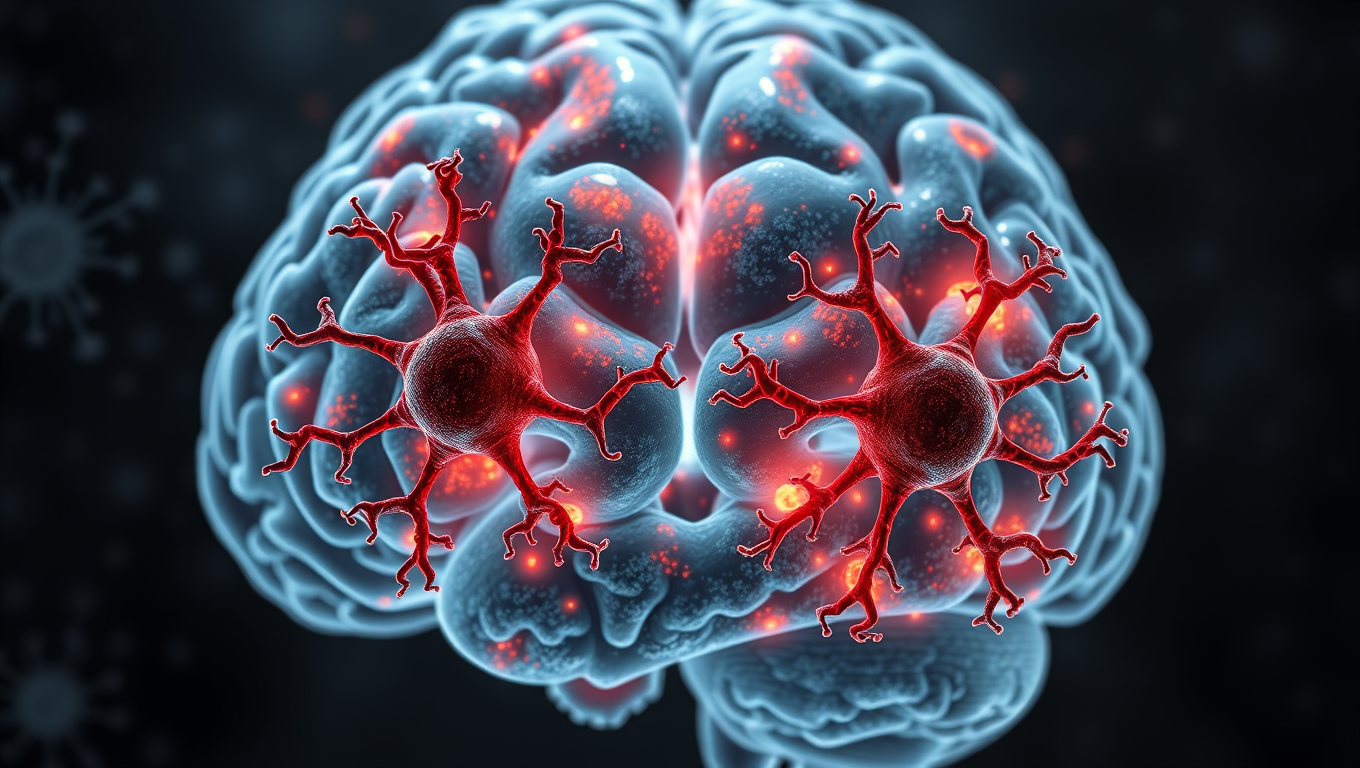
Breaking New Ground: Immune System Discovery Offers Potential Solution to Alzheimer’s
A new way of thinking about Alzheimer’s disease has yielded a discovery that could be the key to stopping the cognitive decline seen in Alzheimer’s and other neurodegenerative diseases, including ALS and Parkinson’s.

Alzheimer's
Different Versions of APOE Protein Alter Microglia Function in Alzheimer’s Disease
A new study suggests how APOE2 is protective while APOE4 increases disease risk by regulating the brain’s immune cells.

A New Horizon for Vision: How Gold Nanoparticles May Restore People’s Sight

Revolutionizing Quantum Communication: Direct Connections Between Multiple Processors

Retiring Abroad Can Be Lonely Business
Harnessing Water Waves: A Breakthrough in Controlling Floating Objects

“Unveiling Hidden Patterns: A New Twist on Interference Phenomena”

Household Electricity Three Times More Expensive Than Upcoming ‘Eco-Friendly’ Aviation E-Fuels, Study Reveals
Reducing Falls Among Elderly Women with Polypharmacy through Exercise Intervention
-
 Detectors1 year ago
A New Horizon for Vision: How Gold Nanoparticles May Restore People’s Sight
-
Cancer1 year ago
Revolutionizing Quantum Communication: Direct Connections Between Multiple Processors
-
Earth & Climate1 year ago
Retiring Abroad Can Be Lonely Business
-
Albert Einstein1 year ago
Harnessing Water Waves: A Breakthrough in Controlling Floating Objects
-
Chemistry1 year ago
“Unveiling Hidden Patterns: A New Twist on Interference Phenomena”
-
Earth & Climate1 year ago
Household Electricity Three Times More Expensive Than Upcoming ‘Eco-Friendly’ Aviation E-Fuels, Study Reveals
-
Diseases and Conditions1 year ago
Reducing Falls Among Elderly Women with Polypharmacy through Exercise Intervention
-
Acid Rain1 year ago
“Revolutionizing Honey Bee Survival: A New Pollen-Replacing Food Source Brings Hope for the Future”