Connect with us

Biochemistry

Breaking Ground: Terahertz Imaging Revolutionizes Cochlear Visualization

Researchers have discovered a groundbreaking use of terahertz (THz) imaging to visualize cochlear structures in mice, offering non-invasive, high-resolution diagnostics. By creating 3D reconstructions, this technology opens new possibilities for diagnosing hearing loss and other conditions. THz imaging could lead to miniaturized devices, like THz endoscopes and otoscopes, revolutionizing diagnostics for hearing loss, cancer, and more. With the potential to enhance diagnostic speed, accuracy, and patient outcomes, THz imaging could transform medical practices.
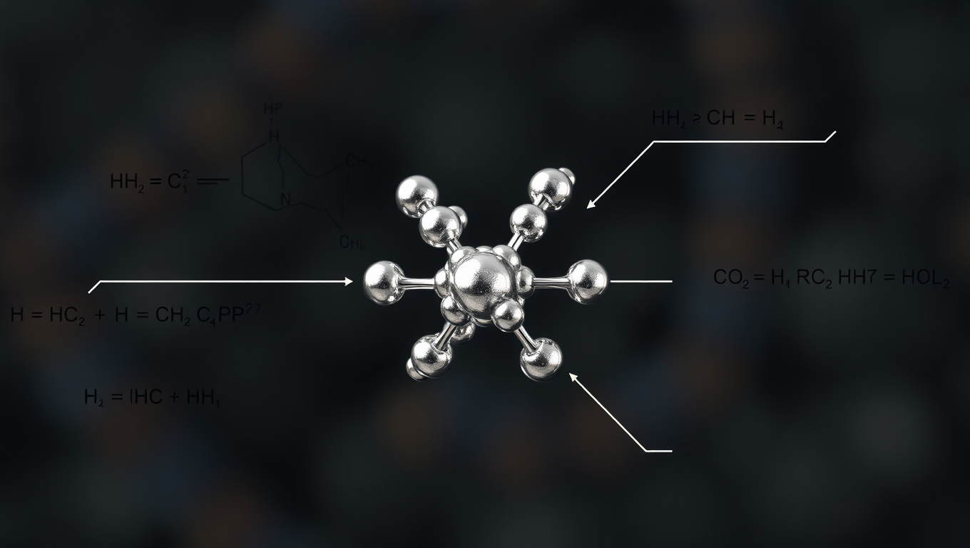
Avatar photo

Published

on

Breaking Ground: Terahertz Imaging Revolutionizes Cochlear Visualization

The world of medical imaging has witnessed a significant breakthrough with the discovery of a groundbreaking use of terahertz (THz) imaging to visualize cochlear structures in mice. This non-invasive, high-resolution diagnostic approach has opened new possibilities for diagnosing hearing loss and other conditions, paving the way for a revolution in medical practices.

The aging population is facing an alarming rise in age-related disorders that significantly impact overall well-being. Hearing loss, in particular, affects communication, social interactions, and daily functioning, making its precise visualization essential for understanding and diagnosing auditory disorders. Conventional imaging techniques often struggle to capture the intricate details of this delicate structure, necessitating the development of more advanced imaging approaches.

Researchers from Waseda University, Kobe University, and Osaka University have successfully demonstrated the first THz imaging of the internal structure of the mouse cochlea using a micrometer-sized THz point source. The study, published in Opticaon 27 March, 2025, explores THz imaging as a non-invasive, high-resolution technique for biological tissue analysis.

The team achieved high-resolution THz imaging by generating a micrometer-sized THz point source using a femtosecond laser at a wavelength of 1.5 μm and irradiating a GaAs substrate. The cochlea was placed directly on the substrate to facilitate near-field imaging, capturing 2D THz time-domain images over a broad timescale. By applying the time-of-flight principle, the time scale of each THz image was converted into a depth scale.

To enable 3D reconstruction of the cochlea, k-means clustering, an unsupervised machine-learning technique, was used to extract structural features. The resulting 3D point cloud and surface mesh model provided high-quality spatial representations of the cochlea, enhancing our understanding of its internal architecture.

The findings of this study highlight the potential of THz imaging as a viable alternative to conventional methods for inner ear diagnostics. The proposed THz imaging technique could be developed into miniaturized devices, such as THz endoscopes and otoscopes, enabling non-invasive, in vivo imaging for cochlear diagnostics, dermatology, and early cancer detection.

The integration of THz technology with existing medical devices holds great potential for revolutionizing the way diseases are diagnosed, particularly in oncology and pathology. “The speed and accuracy of pathological diagnoses could be significantly enhanced by THz technology,” says Associate Professor Kazunori Serita from Waseda University, adding that it may ultimately improve patient outcomes.

By demonstrating the potential of THz imaging for visualizing the cochlea through near-field imaging and 3D reconstruction, this study explores its possible applications in biomedical diagnostics. With its non-invasive, high-resolution capabilities, THz technology may offer a useful approach for medical imaging and analysis, paving the way for significant advancements in medical practices.

Biochemistry

“Tailoring Gene Editing with Machine Learning: A Breakthrough in CRISPR-Cas9 Enzyme Engineering”

Genome editing has advanced at a rapid pace with promising results for treating genetic conditions — but there is always room for improvement. A new paper showcases the power of scalable protein engineering combined with machine learning to boost progress in the field of gene and cell therapy. In their study, authors developed a machine learning algorithm — known as PAMmla — that can predict the properties of about 64 million genome editing enzymes. The work could help reduce off-target effects and improve editing safety, enhance editing efficiency, and enable researchers to predict customized enzymes for new therapeutic targets.

Avatar photo

Published

on

The article “Tailoring Gene Editing with Machine Learning: A Breakthrough in CRISPR-Cas9 Enzyme Engineering” discusses how researchers from Mass General Brigham have harnessed machine learning to revolutionize the field of genome editing. By developing a machine learning algorithm called PAMmla, they’ve predicted the properties of over 64 million genome editing enzymes, significantly expanding our repertoire of effective and safe CRISPR-Cas9 enzymes.

CRISPR-Cas9 enzymes are powerful tools for editing genes, but their traditional application can have off-target effects, modifying DNA at unintended sites in the genome. The researchers’ novel approach uses machine learning to better predict and tailor these enzymes, ensuring greater specificity and accuracy in gene editing. This scalable solution has the potential to transform our understanding of genetic conditions and unlock new therapeutic targets.

The study showcases the power of PAMmla by demonstrating its utility in precise editing disease-causing sequences in primary human cells and mice. The researchers have also made a web tool available for others to use this model, enabling the community to create customized enzymes tailored for specific research and therapeutic applications.

Ben Kleinstiver, PhD, and Rachel A. Silverstein, PhD candidate, are leading authors on this study, highlighting the potential of machine learning in expanding our capabilities in gene editing. This breakthrough has significant implications for the field, offering a new era of precision and safety in genome editing technology.

Continue Reading

Biochemistry

Unlocking Cellular Secrets: New Technique Expands Tissues for Mass Spectrometry Imaging

A new tissue expansion method enables scientists to use mass spectrometry imaging to simultaneously detect hundreds of molecules at the single cell level in their native locations.

Avatar photo

Published

on

Unlocking Cellular Secrets: New Technique Expands Tissues for Mass Spectrometry Imaging

For biologists, seeing is believing. But sometimes biologists face a daunting challenge: visualizing the intricate world within intact tissue samples, down to the level of single cells. Detecting hundreds or thousands of biomolecules – from lipids to metabolites to proteins – in their native environment allows researchers to better understand their functions and interactions.

Traditional imaging methods, including most types of microscopy, provide a view of molecules inside cells but can only track a select handful at one time. Other methods, like regular mass spectrometry, can detect hundreds of molecules but don’t work on intact samples, so researchers can’t see how the biomolecules are oriented.

One promising technique – mass spectrometry imaging – overcomes some of these challenges. It allows researchers to see hundreds of molecules at once in intact tissues. However, it doesn’t have high enough resolution to allow detection at the single cell level.

This was the problem Janelia Senior Group Leader Meng Wang faced. Wang and her team study the fundamental mechanisms behind aging and longevity, and they wanted to detect many different biomolecules in intact tissues to understand how the components change as tissues age.

“Knowing at each specific location what molecules are there and what is in the neighboring cells is very important for any kind of biological question,” Wang says.

Luckily, Wang’s lab is down the hall from Janelia Principal Scientist Paul Tillberg. Tillberg co-invented expansion microscopy as a graduate student at MIT. The method uses a swellable hydrogel material to expand samples uniformly in all directions to a point where fine details, like sub-organelle structure, can be detected with a conventional microscope.

Now a decade old, the expansion process is being applied to other methods outside traditional microscopy. Wang, Tillberg, and their collaborators at Janelia and the University of Wisconsin-Madison wanted to see if they could use expansion to overcome mass spectrometry imaging’s spatial resolution problem.

The result is a new method that expands tissue samples gradually without having to degrade them at the molecular level, as happens in the original expansion process. By expanding the intact samples in all directions, researchers can use mass spectrometry imaging to simultaneously detect hundreds of molecules at the single cell level in their native locations.

“This lets you have an untargeted look in the molecular space, and we are trying to bring it closer to what microscopy can do in terms of spatial resolution,” Tillberg says.

The team used the new technique to delineate the specific spatial patterns of small molecules in different layers of the cerebellum. They found that these molecules – including lipids, peptides, proteins, metabolites, and glycans – are not uniformly distributed, as previously thought. Moreover, they found that each specific layer of the cerebellum has its own signature of lipids, metabolites, and proteins.

The team was also able to detect biomolecules in kidney, pancreas, and tumor tissues, demonstrating that the method can be adapted for many different tissue types. In tumor tissues, they were able to visualize large variations in biomolecules, which could be useful for understanding the molecular mechanisms of tumors and potentially aid in drug development.

“When you can see these biomolecules, then you can start to understand why they have such patterns and how that is related to function,” says Wang. She believes the new technology will allow researchers to track these patterns during development, aging, and disease to understand how different molecules contribute to these processes.

Because the new method doesn’t require adding hardware to an existing mass spec imaging system, and the expansion technique is relatively easy to learn, the team hopes it will be used by many labs around the world. They also hope the new technique will make mass spectrometry imaging a more useful tool for biologists and have laid out a detailed description of the new method and a roadmap for adapting it to other tissue types.

“We wanted to develop something that did not require specialized instruments or procedures, but can be broadly adopted,” Wang says.

Continue Reading

Alternative Fuels

A Breakthrough in Green Hydrogen Production: Cage Structured Material Transforms into High-Performance Catalyst

Clathrates are characterized by a complex cage structure that provides space for guest ions too. Now a team has investigated the suitability of clathrates as catalysts for electrolytic hydrogen production with impressive results: the clathrate sample was even more efficient and robust than currently used nickel-based catalysts. They also found a reason for this enhanced performance. Measurements at BESSY II showed that the clathrates undergo structural changes during the catalytic reaction: the three-dimensional cage structure decays into ultra-thin nanosheets that allow maximum contact with active catalytic centers.

Avatar photo

Published

on

Rewritten Article:

Scientists have made a groundbreaking discovery that could revolutionize the production of green hydrogen, a crucial component for a sustainable energy future. Researchers have found that a cage-structured material, previously unknown as an electrocatalyst, can outperform existing nickel-based catalysts in electrolytic hydrogen production. This breakthrough has significant implications for the chemical industry and our transition to renewable energy sources.

The study, published in Angewandte Chemie, investigates the suitability of clathrates – materials characterized by a complex three-dimensional cage structure – as catalysts for oxygen evolution reaction (OER) in electrolysis. Clathrates have shown promise in various applications, such as thermoelectrics and superconductors, but their potential as electrocatalysts has remained unexplored until now.

Dr. Prashanth Menezes and his team at the Technical University of Munich synthesized Ba₈Ni₆Ge₄₀ clathrates, which they then tested as OER catalysts in aqueous electrolytes. The results were astonishing: the clathrate sample exceeded the efficiency of nickel-based catalysts at a current density of 550 mA cm⁻², a value commonly used in industrial electrolysis. Moreover, its stability was remarkable, with activity remaining high even after 10 days of continuous operation.

To understand why this material performed so well, the researchers employed a combination of experiments, including in situ X-ray absorption spectroscopy (XAS) at BESSY II and basic structural characterization at the Freie and Technische Universität Berlin. Their analysis revealed that the clathrate particles undergo a structural transformation under an electric field: germanium and barium atoms dissolve out of the former three-dimensional framework, leaving behind highly porous, sponge-like nanolayers of nickel that offer maximum surface area.

“This transformation brings more and more catalytically active nickel centres into contact with the electrolyte,” says Dr. Niklas Hausmann from Menezes’ team. “We were actually surprised by how well these samples work as OER catalysts. We expect that we can observe similar results with other transition metal clathrates and that we have discovered a very interesting class of materials for electrocatalysts.”

This breakthrough has significant implications for the production of green hydrogen, which is seen as an essential building block for a sustainable energy future. With this new material, researchers may be able to develop more efficient and robust OER catalysts, enabling faster and more cost-effective production of green hydrogen.

Continue Reading

Trending