Connect with us

Brain Tumor

Hair-like Electrodes Revolutionize Brain Activity Monitoring

Researchers have created a hairlike device for long-term, non-invasive monitoring of the brain’s electrical activity. The lightweight and flexible electrode attaches directly to the scalp and delivers stable, high-quality electroencephalography (EEG) recordings.
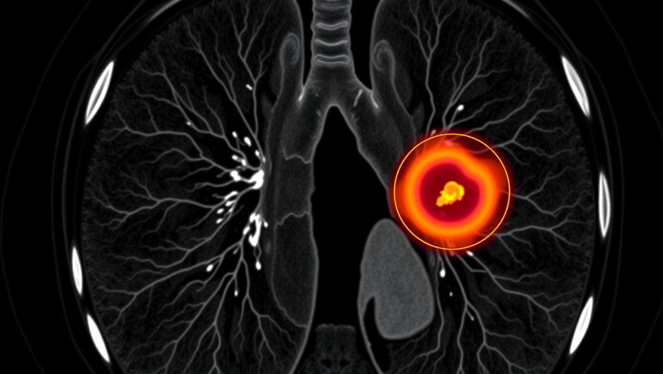
Avatar photo

Published

on

The article you provided has been rewritten for clarity, structure, and style, making it accessible to a general audience. Here’s the rewritten content:

In the realm of electroencephalography (EEG) monitoring, researchers at Penn State have made a groundbreaking discovery – one that could revolutionize the way we monitor brain activity. Gone are the days of cumbersome metal electrodes; instead, a team of scientists has created hair-like devices for non-invasive, long-term monitoring.

The innovative electrode is designed to mimic human hair and can be worn without drawing attention. This lightweight and flexible device captures stable, high-quality recordings of the brain’s signals for over 24 hours of continuous wear. The traditional metal electrodes used in EEG monitoring are rigid and can shift when someone moves their head, compromising data uniformity.

The new electrode uses a 3D-printed bioadhesive ink that allows it to stick directly onto the scalp without any gloopy gels or skin preparation. This minimizes the gap between the electrode and skin, improving signal quality. The device is also stretchable, ensuring it stays put even when combing hair or wearing a baseball cap.

The researchers found that the new device performed comparably to gold electrodes, the current standard for EEG monitoring. However, the hair-like electrode maintained better contact between the electrode and skin and performed reliably for extended periods without any degradation in signal quality.

According to Tao Zhou, Wormley Family Early Career Professor of Engineering Science and Mechanics, this technology holds promise for use in consumer health and wellness products, as well as clinical healthcare applications.

The conventional EEG monitoring process can be a cumbersome affair, requiring the application of gels to maintain good surface-to-surface contact between the electrodes and skin. This process is imprecise and can result in different amounts of gel used on the electrodes, affecting brain signal quality.

Zhou explained that this new device will change the impedance – or interface – between the electrodes and scalp, ensuring more consistent and reliable monitoring of EEG signals. The researchers also hope to make the system wireless in the future, allowing people to move around freely during recording sessions.

The team’s findings were published in a study in npc biomedical innovations, with funding from various institutions, including the National Institutes of Health and Oak Ridge Associated Universities.

In conclusion, the development of hair-like electrodes for brain activity monitoring is a significant breakthrough that could revolutionize the field. With its potential for non-invasive, long-term monitoring, this technology has far-reaching implications for healthcare and consumer products alike.

Biochemistry

A Breakthrough in Brain Research: The Iontronic Pipette Revolutionizes Neurological Studies

Researchers have developed a new type of pipette that can deliver ions to individual neurons without affecting the sensitive extracellular milieu. Controlling the concentration of different ions can provide important insights into how individual brain cells are affected, and how cells work together. The pipette could also be used for treatments.

Avatar photo

Published

on

The development of an iontronic pipette at Linköping University has opened up new avenues for neurological research. This innovative tool allows researchers to deliver ions directly to individual neurons without affecting the surrounding extracellular milieu. By controlling the concentration of various ions, scientists can gain valuable insights into how brain cells respond to different stimuli and interact with each other.

The human brain consists of approximately 85-100 billion neurons, supported by a similar number of glial cells that provide essential functions such as nutrition, oxygenation, and healing. The extracellular milieu, a fluid-filled space between the cells, plays a crucial role in maintaining cell function. Changes in ion concentration within this environment can activate or inhibit neuronal activity, making it essential to study how local changes affect individual brain cells.

Previous attempts to manipulate the extracellular environment involved pumping liquid into the area, disrupting the delicate biochemical balance and making it difficult to determine whether the substances themselves or the changed pressure were responsible for the observed effects. To overcome this challenge, researchers at the Laboratory of Organic Electronics developed an iontronic micropipette measuring only 2 micrometers in diameter.

This tiny pipette can deliver ions such as potassium and sodium directly into the extracellular milieu, allowing scientists to study how individual neurons respond to these changes. Glial cell activity is also monitored, providing a more comprehensive understanding of brain function.

Theresia Arbring Sjöström, an assistant professor at LOE, highlighted that glial cells are critical components of the brain’s chemical environment and can be precisely activated using this technology. In experiments conducted on mouse hippocampus tissue slices, it was observed that neurons responded dynamically to changes in ion concentration only after glial cell activity had saturated.

This research has significant implications for neurological disease treatment. The iontronic pipette could potentially be used to develop extremely precise treatments for conditions such as epilepsy, where brain function can be disrupted by localized imbalances in ion concentrations.

Researchers are now continuing their studies on chemical signaling in healthy and diseased brain tissue using the iontronic pipette. They also aim to adapt this technology to deliver medical drugs directly to affected areas of the brain, paving the way for more targeted treatments for neurological disorders.

Continue Reading

Brain Injury

Uncovering the Aggressive Nature of Glioblastoma: ZIP4’s Role in Brain Tumor Growth

Researchers detail their discoveries about why the brain tumor glioblastoma is so aggressive. Their findings center on ZIP4, a protein that transports zinc throughout the body and sets off a cascade of events that drive tumor growth.

Avatar photo

Published

on

In a groundbreaking study published in the Proceedings of the National Academy of Sciences (PNAS), University of Oklahoma researchers have made a significant discovery about what makes glioblastoma, the deadliest form of brain cancer, so aggressive. The findings center on ZIP4, a protein that transports zinc throughout the body and sets off a chain reaction that drives tumor growth.

Glioblastomas account for about half of all malignant brain tumors, with a median survival rate of 14 months. Surgery is often challenging, and patients almost always experience a relapse. By better understanding why these brain tumors are so aggressive, researchers hope to open up paths for new treatments.

In normal conditions, ZIP4 plays a positive role, transporting and maintaining the right amount of zinc for good health. However, when brain cancer is present, ZIP4 takes on a different role. In the case of glioblastoma, it triggers a series of events that contribute to the tumor’s aggressive growth.

“Everything starts with the fact that ZIP4 is overexpressed in glioblastoma,” says senior author Min Li, Ph.D., a professor of medicine, surgery, and cell biology at the University of Oklahoma College of Medicine. “That triggers all these downstream events that help the tumor to grow.”

Li’s research team tested a small-molecule inhibitor that targets ZIP4 and TREM1, a protein involved in immune responses. The inhibitor attached to both proteins, stopping their actions and slowing tumor growth. This suggests that ZIP4 and TREM1 may be promising therapeutic targets.

Neurosurgeon Ian Dunn, M.D., executive dean of the OU College of Medicine and co-author of the study, says the findings are an encouraging step toward combating this debilitating cancer. “These results are really exciting in such a debilitating cancer. The hope and promise is to translate these findings to novel treatment approaches to improve the lives of our patients.”

This discovery is significant not only for glioblastoma but also for pancreatic cancer research, as ZIP4 has been a focus of Li’s work on this disease for many years. He found that overexpression of ZIP4 causes pancreatic cancer cells to be more resistant to chemotherapy and prompts tumor cells to transform themselves so they can stealthily travel to the body’s other organs.

The researchers hope that their findings will lead to new treatment approaches for glioblastoma and potentially other types of cancer, improving the lives of patients affected by these devastating diseases.

Continue Reading

Alzheimer's

“Unlocking Brain Resilience: Researchers Discover CLU Protein’s Potential to Protect Against Alzheimer’s Disease”

Recently approved Alzheimer’s drugs offer a step forward for treating the disease, but new therapeutic strategies are needed to complement them and provide personalized therapeutic approaches. Researchers have identified a potential strategy to protect against Alzheimer’s disease by increasing the clusterin protein (CLU).

Avatar photo

Published

on

As scientists continue to search for effective treatments for Alzheimer’s disease, a recent study has shed light on a promising new approach. Researchers at Mass General Brigham have discovered that increasing levels of the “CLU” (clusterin) protein may protect against cognitive decline and potentially provide a complementary therapy to existing treatments.

Led by Dr. Tracy Young-Pearse, the research team used various models, including human brain tissue from over 700 participants and animal models, to uncover the molecular role of CLU in protecting against neurodegeneration. Their findings suggest that increasing CLU can prevent inflammatory interactions between brain cells called astrocytes and microglia, which are associated with heightened inflammation.

The study’s results have significant implications for the design and testing of new therapeutic strategies for Alzheimer’s disease. As Dr. Young-Pearse noted, “Increasing clusterin has the potential to prevent cognitive decline in a way that is different than and complementary to anti-amyloid therapies to promote brain resilience.”

CLU may also be beneficial for treating other age-related brain diseases, many of which share similar mechanisms of neuroinflammatory dysregulation. The researchers emphasize the importance of personalized therapeutic approaches, as individuals’ responses to CLU upregulation vary based on their genetics.

The study’s findings provide a crucial step forward in the search for effective treatments for Alzheimer’s disease and other age-related brain diseases. By understanding the role of CLU in protecting against neurodegeneration, researchers can develop new strategies to promote brain resilience and potentially prevent cognitive decline.

In conclusion, the discovery of CLU’s potential therapeutic benefits offers new hope for individuals affected by Alzheimer’s disease and other age-related brain disorders. Further research is necessary to fully explore this promising approach, but the findings of this study mark an important milestone in the pursuit of effective treatments for these debilitating diseases.

Continue Reading

Trending