Connect with us
We’re experimenting with AI-generated content to help deliver information faster and more efficiently.
While we try to keep things accurate, this content is part of an ongoing experiment and may not always be reliable.
Please double-check important details — we’re not responsible for how the information is used.

Diseases and Conditions

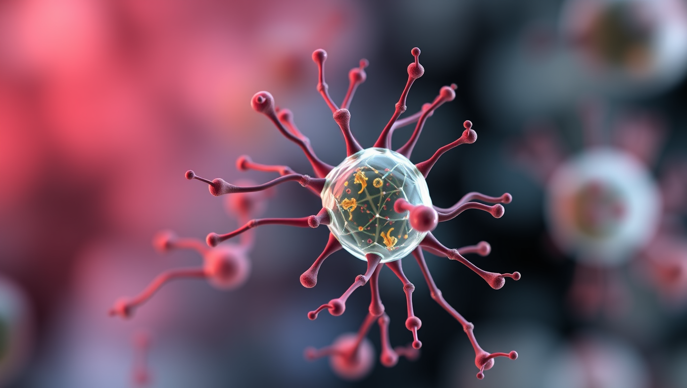
“Revolutionizing Cancer Treatment: AI-Powered Model Predicts Cell Activity in Tissues Over Time”

A team of scientists has developed a remarkable new approach to modeling how cells behave over time—using a digital “forecast” much like predicting the weather. By combining patient genomics with a groundbreaking plain-language “hypothesis grammar,” the researchers can simulate how cells communicate and evolve within tissues. These simulations allow scientists to digitally test how cancers grow, how immune systems respond, and even how treatments might work in individual patients.

Avatar photo

Published

on

The new software combines genomics technologies with computational modeling to predict cell changes in behavior, such as communication between cells that could cause cancer cells to flourish.

Researchers at the University of Maryland School of Medicine’s Institute for Genome Sciences (IGS) co-led the study that published online on July 25 in the journal Cell. It is the result of a multi-year, multi-lab project at the interface of software development with important collaborations between bench and clinical team science researchers.

“This research eventually could lead to computer programs that could help determine the best treatment for cancer patients by essentially creating a ‘digital twin’ of the patient,” said Jeanette Johnson, PhD, a Postdoc Fellow at the Institute for Genome Sciences (IGS) at UMSOM and co-first author of this study.

The team used a plain-language “hypothesis grammar” that uses common language as a bridge between biological systems and computational models to simulate how cells act in tissue. This grammar allows scientists to use simple English language sentences to build digital representations of multicellular biological systems and enabled the team to develop computational models for diseases like cancer.

In breast cancer, the IGS team modeled an effect where the immune system cannot curtail tumor cell growth and instead promotes invasion and cancer spread. They adapted this computational modeling framework to simulate a real-world immunotherapy clinical trial of pancreatic cancer.

Using genomics data from untreated tissue samples of pancreatic cancer, the model predicted that each virtual “patient” had a different response to the immunotherapy treatment — showcasing the importance of cellular ecosystems for precision oncology.

The team used new spatial genomics technology to further demonstrate the ways fibroblasts communicate with tumor cells. The program allowed the scientists to follow the growth and progression of pancreatic tumors to invasion from real patient tissue.

“What makes these models so exciting to me as someone who studies immunology is that they can be informed, initialized, and built upon using both laboratory and human genomics data,” said Dr. Johnson.

The new grammar is open source so that all scientists can benefit from it. “By making this tool accessible to the scientific community, we are providing a path forward to standardize such models and make them generally accepted,” said Daniel Bergman, PhD, a scientist at IGS and Assistant Professor of Pharmacology and Physiology at UMSOM.

To demonstrate this generalizability, Genevieve Stein-O’Brien, PhD, the Terkowitz Family Rising Professor of Neuroscience and Neurology at Johns Hopkins School of Medicine (JHSOM) led researchers in using this approach in a neuroscience example in which the program simulated the creation of layers as the brain develops.

“With this work from IGS, we have a new framework for biological research since researchers can now create computerized simulations of their bench experiments and clinical trials and even start predicting the effects of therapies on patients,” said Mark T. Gladwin, MD, Vice President for Medical Affairs at the University of Maryland, Baltimore, and the John Z. and Akiko K. Bowers Distinguished Professor and UMSOM Dean.

The team of senior authors on this study include, Paul Macklin, PhD, Associate Dean for Undergraduate Education and Professor of Intelligent Systems Engineering at the Indiana School of Informatics, Computing and Engineering at Indiana University, Genevieve Stein-O’Brien, Bloomberg Assistant Professor of Neuroscience and Assistant Director Single-Cell Training and Analysis Center (STAC) at Johns Hopkins University, and Dr. Fertig are continuing efforts to disseminate this software and extend its integration with genomics data for automatic model formulation through the National Cancer Institute (NCI) Informatics Technology in Cancer Research Consortium, who funded this study. Additional benchmarking of this study and applications of the software to breast and pancreatic cancer are supported from numerous NCI grants, the Jayne Koskinas Ted Giovanis Foundation, the National Foundation for Cancer Research, the Cigarette Restitution Fund Program from the State of Maryland, and the Lustgarten Foundation.

Birth Control

A Safer, Cheaper Vision Correction Method May Be on the Horizon

Scientists are developing a surgery-free alternative to LASIK that reshapes the cornea using electricity instead of lasers. In rabbit tests, the method corrected vision in minutes without incisions.

Avatar photo

Published

on

This revolutionary approach to vision correction is called electromechanical reshaping (EMR). This method utilizes electrical impulses to reshape the cornea, potentially providing a safer and more affordable alternative to traditional LASIK surgery. Researchers at Occidental College have made significant progress in this area, with initial studies demonstrating promising results on rabbit eyeballs.

The researchers’ innovative technique involves using platinum “contact lenses” that provide a template for the corrected shape of the cornea. By applying an electric potential to these contact lenses, they create a precise pH change within the tissue, loosening its rigidity and making it malleable. This enables them to reshape the cornea without any incisions or ablative procedures.

In their experiments on rabbit eyeballs, the team successfully reshaped the corneas of 12 separate specimens, 10 of which were treated as if they had myopia (nearsightedness). The treatment effectively corrected the focusing power of the eye in all “myopic” eyeballs. Moreover, the cells within the eyeball survived this procedure because the researchers carefully controlled the pH gradient.

The researchers emphasize that while these initial results are promising, their work is still in its early stages. They plan to conduct further animal studies and investigate the potential of EMR for treating a range of vision problems, including astigmatism, near- and far-sightedness. However, the team’s scientific funding uncertainties have put them on hold.

Despite these challenges, the researchers remain optimistic about the potential of this new technique. They believe that if successful, EMR could provide a widely applicable, vastly cheaper, and potentially even reversible method for vision correction, surpassing current treatments like LASIK.

Continue Reading

Children's Health

Uncovering the Inaccuracy: Why Common Blood Pressure Readings May Miss 30% of Hypertension Cases

Cambridge scientists have cracked the mystery of why cuff-based blood pressure monitors often give inaccurate readings, missing up to 30% of high blood pressure cases. By building a physical model that replicates real artery behavior, they discovered that low pressure below the cuff delays artery reopening, leading to underestimated systolic readings. Their work suggests that simple tweaks, like raising the arm before testing, could dramatically improve accuracy without the need for expensive new devices.

Avatar photo

Published

on

The measurement of blood pressure has been a cornerstone of medical practice for decades. However, despite its widespread use, research suggests that common cuff-based blood pressure readings may be inaccurate and potentially miss up to 30% of hypertension cases.

A team of researchers from the University of Cambridge has shed new light on this issue by building an experimental model that explains the physics behind these inaccuracies. Their findings, reported in the journal PNAS Nexus, have significant implications for patient health outcomes and highlight the need for more accurate measurement methods.

The auscultatory method, which relies on inflating a cuff around the upper arm to measure blood pressure, has long been considered the gold standard. However, this study reveals that it overestimates diastolic pressure while underestimating systolic pressure. The researchers attribute this discrepancy to a previously unidentified factor: the delayed reopening of arteries due to low downstream pressure.

To replicate this condition in their experimental rig, the Cambridge team used tubes that lay flat when deflated and fully closed when inflated with cuff pressure. This setup allowed them to study the effects of downstream blood pressure on artery closure and reopening, leading to a better understanding of the mechanics behind inaccurate readings.

The researchers propose several potential solutions to address this underestimation, including raising the arm before measurement to produce a predictable downstream pressure. This simple change does not require new devices but can make blood pressure measurements more accurate.

If new devices for monitoring blood pressure are developed, they may incorporate additional inputs that correlate with downstream pressure, such as age, BMI, or tissue characteristics, to adjust ‘ideal’ readings for each individual.

The study’s authors emphasize the need for clinical trials to test their findings in patients and collaborate with clinicians to implement changes to clinical practice. Funding from organizations like the Engineering and Physical Sciences Research Council (EPSRC) will be essential to support further research and development.

By uncovering the inaccuracies in common blood pressure readings, this study has significant implications for patient health outcomes and highlights the need for more accurate measurement methods. The proposed solutions have the potential to improve diagnosis and treatment of hypertension, ultimately saving lives and reducing healthcare costs.

Continue Reading

Allergy

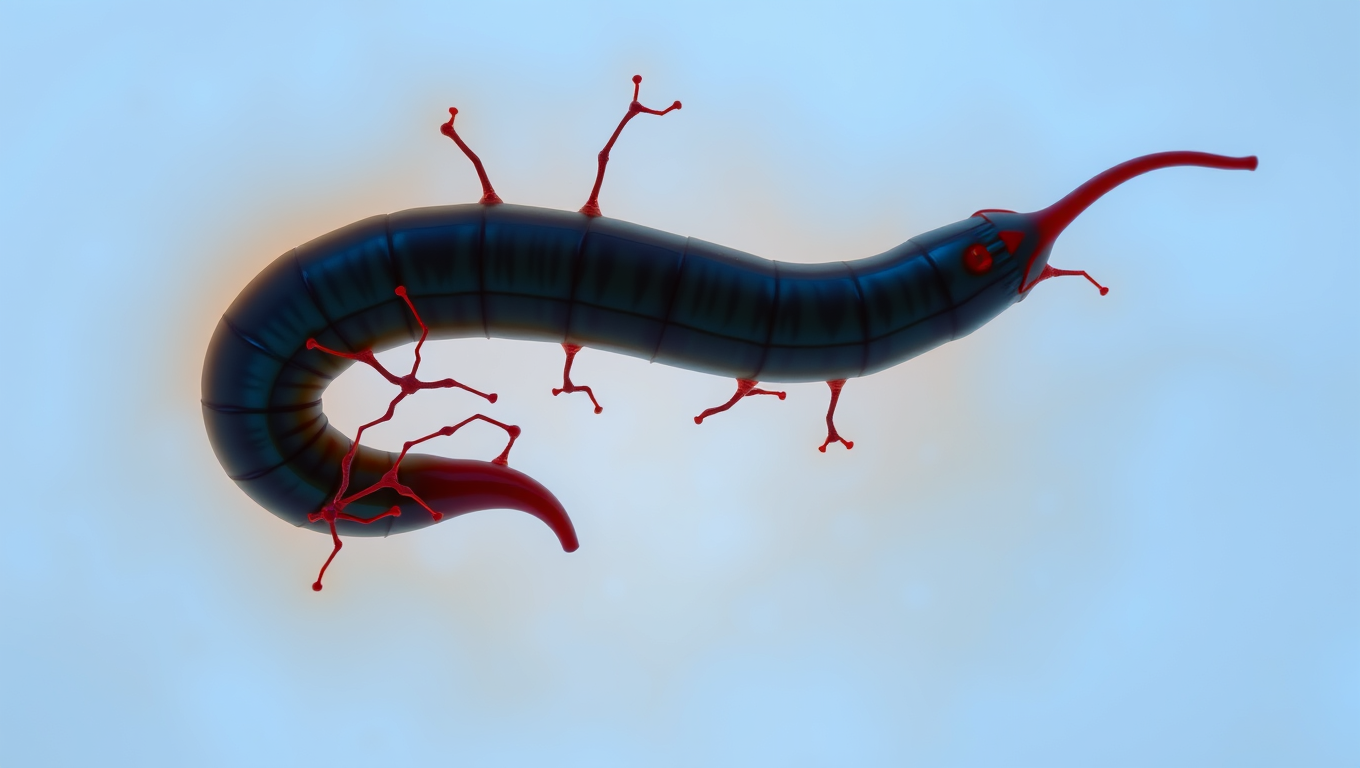
“The Silent Invader: How a Parasitic Worm Evades Detection and What it Can Teach Us About Pain Relief”

Scientists have discovered a parasite that can sneak into your skin without you feeling a thing. The worm, Schistosoma mansoni, has evolved a way to switch off the body’s pain and itch signals, letting it invade undetected. By blocking certain nerve pathways, it avoids triggering the immune system’s alarms. This stealth tactic not only helps the worm survive, but could inspire new kinds of pain treatments and even preventative creams to protect people from infection.

Avatar photo

Published

on

A new study published in The Journal of Immunology has made an intriguing discovery about how a parasitic worm evades detection and what it can teach us about pain relief. Researchers from Tulane School of Medicine found that the Schistosoma mansoni worm, which causes schistosomiasis, suppresses neurons in the skin to avoid triggering an immune response.

When this worm penetrates human skin, typically through contact with infested water, it produces molecules that block a protein called TRPV1+, which is responsible for sending pain signals to the brain. This clever mechanism allows the worm to infect the skin largely undetected.

The researchers believe that the S. mansoni worm evolved this strategy to enhance its own survival and found that blocking TRPV1+ also reduced disease severity in mice infected with the parasite. The study suggests that identifying the molecules responsible for suppressing TRPV1+ could lead to new painkillers that do not rely on opioids.

Moreover, the researchers discovered that TRPV1+ is essential for initiating host protection against S. mansoni infection. When this protein is activated, it triggers a rapid mobilization of immune cells, which induces inflammation and helps fight off the parasite. This finding highlights the critical role of neurons in pain-sensing and immune responses.

The study’s lead author, Dr. De’Broski R. Herbert, emphasizes that identifying these molecules could inform preventive treatments for schistosomiasis. He envisions a topical agent that activates TRPV1+ to prevent infection from contaminated water for individuals at risk of acquiring S. mansoni.

This groundbreaking research has the potential to revolutionize our understanding of pain relief and immune responses, offering new avenues for developing innovative therapies that could benefit millions worldwide.

Continue Reading

Trending