Connect with us

Brain Injury

The Brain’s Word Memory Centers: Uncovering the Secrets of Language Recall

The parts of the brain that are needed to remember words, and how these are affected by a common form of epilepsy, have been identified by a team of neurologists and neurosurgeons at UCL.
Avatar photo

Published

on

The recent study by neurologists and neurosurgeons at UCL has shed light on the parts of the brain responsible for remembering words. The research, published in Brain Communications, reveals that shrinkage in specific areas of the front and side of the brain (prefrontal, temporal, and cingulate cortices, and the hippocampus) is linked to difficulty recalling words.

This groundbreaking discovery highlights the dispersed network involved in creating and storing word memories throughout the brain. The findings are crucial for understanding conditions such as epilepsy, where patients often struggle with remembering words. The researchers hope that their work will guide neurosurgical treatment for epilepsy patients by helping surgeons avoid damaging areas of the brain essential for language and memory during operations.

Corresponding author, Professor John Duncan (UCL Queen Square Institute of Neurology), emphasized the importance of word recall in daily life: “Being able to remember and recall words is vital for day-to-day memory to function well.” He added that detailed MRI scans can help identify causes of epilepsy and show if any parts of the brain are shrunken, allowing researchers to determine which areas are used for making and storing memories.

In a first-of-its-kind study, the team examined 84 people with temporal lobe epilepsy (epilepsy arising from the temporal lobe at the sides of the head) and hippocampal sclerosis (a condition in which part of the brain is scarred, affecting memory), as well as 43 healthy individuals. High-resolution MRI scans were used to measure the size and shape of different parts of the brain, including the cerebral cortex (the outer layer responsible for thinking, memory, attention, perception, awareness, and language) and specific areas within the hippocampus.

The researchers found that smaller sizes in certain brain areas, like the prefrontal, temporal, and cingulate cortices, and parts of the hippocampus, were linked to worse memory for words in people with epilepsy arising from their temporal lobes. These findings are significant not only for understanding conditions such as epilepsy but also for understanding how the brain organizes and remembers words.

Lead author, Dr Giorgio Fiore (National Hospital for Neurology and Neurosurgery, UCLH), noted that this research is essential for designing neurosurgical operations for epilepsy that will not worsen memory. The work was funded by Epilepsy Research UK and supported by the National Institute for Health and Care Research University College London Hospitals Biomedical Research Centre (NIHR UCLH BRC).

Brain Injury

Favorite Music Triggers a Natural High: Uncovering the Brain’s Secret to Musical Pleasure

A new imaging study showed that listening to favorite music affects the function of the brain’s opioid system.

Avatar photo

Published

on

Favorite music has long been known to evoke intense pleasure, often accompanied by physical sensations such as pleasant “chills.” However, the brain mechanisms behind this phenomenon have remained somewhat of a mystery. Recently, a groundbreaking study conducted at the Turku PET Centre in Finland shed new light on this topic, revealing that listening to favorite music activates the brain’s opioid system.

The study utilized positron emission tomography (PET) and functional magnetic resonance imaging (fMRI) to examine how the brain responds to musical enjoyment. Participants were asked to listen to their favorite music while undergoing these scans, which measured the release of opioids in the brain as well as the density of opioid receptors.

The results showed that listening to favorite music influenced opioid release in several brain areas associated with pleasure, including those linked to the experience of pleasurable chills. Moreover, individual differences in opioid receptor density were found to correlate with brain activation during music listening – the more receptors participants had, the stronger their brains reacted.

According to Academy Research Fellow Vesa Putkinen from the University of Turku, “These results show for the first time directly that listening to music activates the brain’s opioid system. The release of opioids explains why music can produce such strong feelings of pleasure, even though it is not a primary reward necessary for survival or reproduction.”

This study provides significant new insight into how the brain’s chemical systems regulate musical pleasure and may also have practical implications for pain management and mental health treatment. As Professor Lauri Nummenmaa notes, “The brain’s opioid system is involved in pain relief. Based on our findings, the previously observed pain-relieving effects of music may be due to music-induced opioid responses in the brain.”

This rewritten article maintains the core ideas of the original but presents them in a clearer and more engaging manner, making it accessible to a general audience. The prompt for image generation is designed to visually represent the concept of musical pleasure and its neural mechanisms, further emphasizing the key findings of the study.

Continue Reading

Birth Defects

Breaking Down Barriers: Towards Gene-Targeting Drugs for Brain Diseases

Getting therapeutic drugs past the blood-brain barrier has long been a major challenge in treating brain diseases. Now, researchers have explored how cholesterol-modified heteroduplex oligonucleotides (Chol-HDOs) enhance drug delivery to the brain. Their study reveals that Chol-HDOs bind tightly to serum proteins, allowing them to persist in the bloodstream and cross into brain tissue. These findings offer insights into gene-targeting therapies and could help develop treatments for conditions like Alzheimer’s disease.

Avatar photo

Published

on

Breaking Down Barriers: Towards Gene-Targeting Drugs for Brain Diseases

The human brain is a complex and intricate organ that has long been a challenge to treat when it comes to diseases like Alzheimer’s, Parkinson’s, and brain cancers. One of the major obstacles in delivering therapeutic drugs to the brain is the blood-brain barrier (BBB), a protective layer that restricts the passage of molecules from the bloodstream into the brain.

To overcome this hurdle, researchers at Tokyo University of Science have been exploring new ways to deliver gene-targeting drugs, specifically antisense oligonucleotides (ASOs) and heteroduplex oligonucleotides (HDOs), directly to the brain. In a recent study published in the Journal of Controlled Release, the team led by Professor Makiya Nishikawa demonstrated that modifying HDOs with cholesterol molecules (Chol-HDOs) could improve their stability and specificity, allowing them to penetrate the cerebral cortex beyond the blood vessels.

The key to this success lies in how Chol-HDOs interact with proteins in the bloodstream. Unlike ASOs and HDOs, which bind electrostatically to serum proteins with low affinity and are taken up by cells, Chol-HDOs bind tightly to serum proteins, including lipoproteins, via hydrophobic interactions. This strong binding results in slow clearance from the bloodstream, allowing Chol-HDOs to remain in circulation for a longer period.

The researchers also showed that inhibiting scavenger receptors in cells reduces the uptake of both ASOs and Chol-HDOs in the liver and kidneys, shedding light on how these compounds are taken up by different organs. This finding has significant implications for the design of brain-targeting drugs based on Chol-HDOs.

With over 55 million people living with dementia worldwide and 300,000 cases of brain cancer reported annually, the potential therapeutic applications of modified HDOs are vast. The possibility of efficiently delivering ASOs and other nucleic acid-based drugs to the brain may lead to the development of treatments for brain diseases with significant unmet medical needs.

This study provides valuable insight into how brain-targeting drugs could be designed based on Chol-HDOs, paving the way for a new generation of compounds that effectively target brain diseases. As research continues, we can expect modified HDOs to offer hope to millions of patients and their families around the world.

Continue Reading

Brain Injury

The Lasting Effects of Climate Trauma on Decision-Making

A new study suggests that climate trauma — such as experiencing a devastating wildfire — can have lasting effects on cognitive function.

Avatar photo

Published

on

The Lasting Effects of Climate Trauma on Decision-Making

A recent study from University of California San Diego has shed light on the profound impact that climate trauma can have on an individual’s ability to make decisions. The research, which focused on survivors of the devastating 2018 Camp Fire in Northern California, found that those directly exposed to the disaster struggled with making choices that prioritized long-term benefits.

The study, published in Scientific Reports, provides new evidence that cognitive function – particularly decision-making – is also affected by climate trauma. This is in addition to the well-documented physical and mental health impacts of wildfires, which have become increasingly frequent due to climate change.

The research involved 75 participants, divided into three groups: those who survived the Camp Fire, those who were indirectly exposed to the disaster, and a control group with no direct exposure to the fire. All participants underwent a decision-making task with monetary rewards while their brain activity was recorded using Electroencephalogram (EEG) scans.

The results showed that wildfire survivors were significantly less likely to stick with choices that offered long-term rewards, a behavior tracked by a choice metric known as “Win-Stay.” This suggests that climate trauma can lead to impulsivity and poor decision-making, which can have far-reaching consequences for individuals and communities.

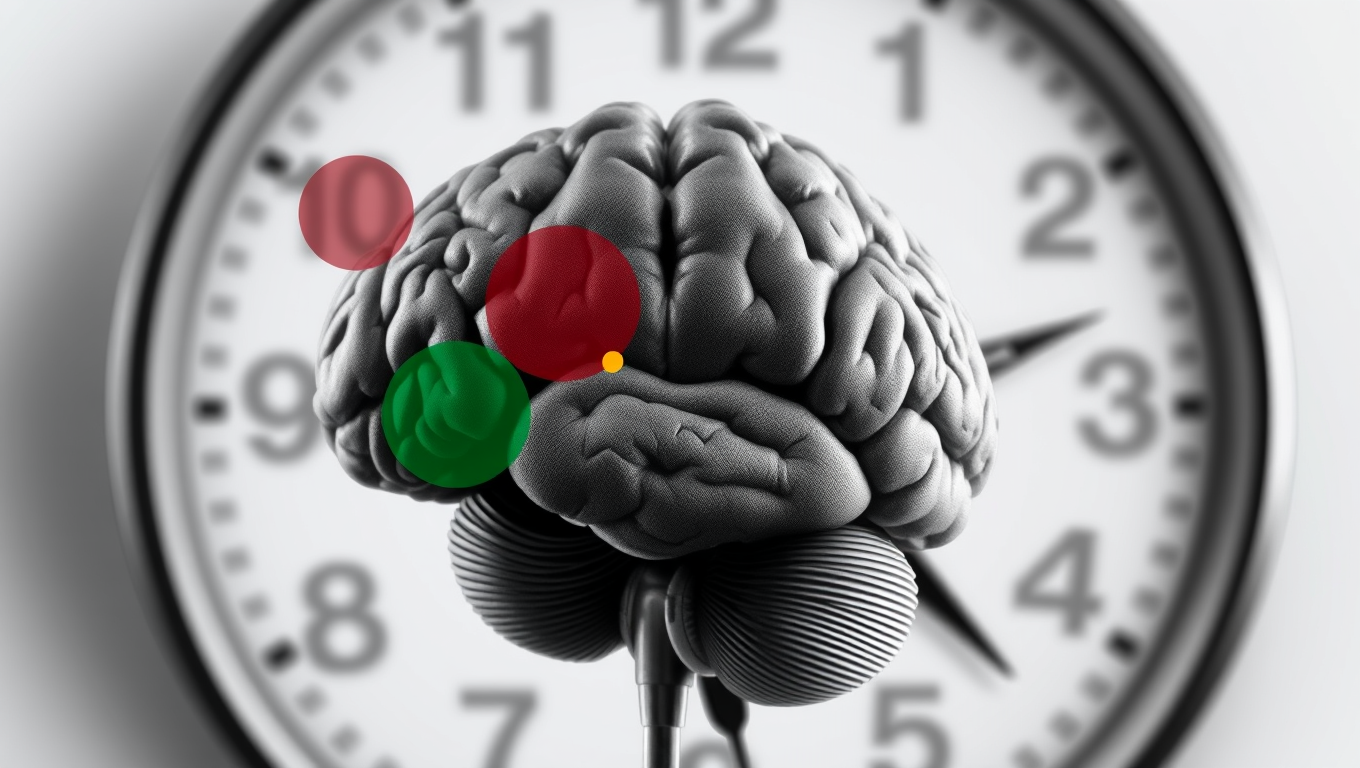
Brain recordings revealed a possible reason why. EEG scans taken while participants engaged in the decision-making task showed heightened activity in the parietal brain region, specifically localized to the posterior cingulate cortex (PCC) – a brain region associated with deep thought and rumination.

“It was clear that brains of study participants directly exposed to wildfires – as opposed to those not exposed – became significantly hyper-aroused when trying to make proper decision choices but they were unable to,” said Jason Nan, a UC San Diego bioengineering graduate student and study first author.

This research has significant implications for the development of new diagnostic tools and personalized treatments for those impacted by climate trauma. One potential intervention is mindfulness and compassion training, which has shown promise in suppressing ruminating thoughts and thereby mitigating the effects of trauma.

As climate disasters become more frequent and severe, researchers emphasize the need to study pre- vs. post-disaster cognitive changes, investigate long-term effects of repeated exposure to climate trauma, and develop scalable mental health interventions for affected communities.

By understanding how climate trauma affects decision-making, we can take steps towards mitigating its impact on individuals and communities, and ultimately build resilience in the face of climate change.

Continue Reading

Trending