While we try to keep things accurate, this content is part of an ongoing experiment and may not always be reliable.
Please double-check important details — we’re not responsible for how the information is used.
Allergy
The Missing Link in Autoimmune Disorders: Researchers Identify Key Protein in Immune Response
Scientists have identified a protein in cells that spurs the release of infection-fighting molecules. The protein, whose role in the immune system had not previously been suspected, provides a potential target for therapies that could prevent over-reactive immune responses that are at the root of several debilitating illnesses.


Allergy
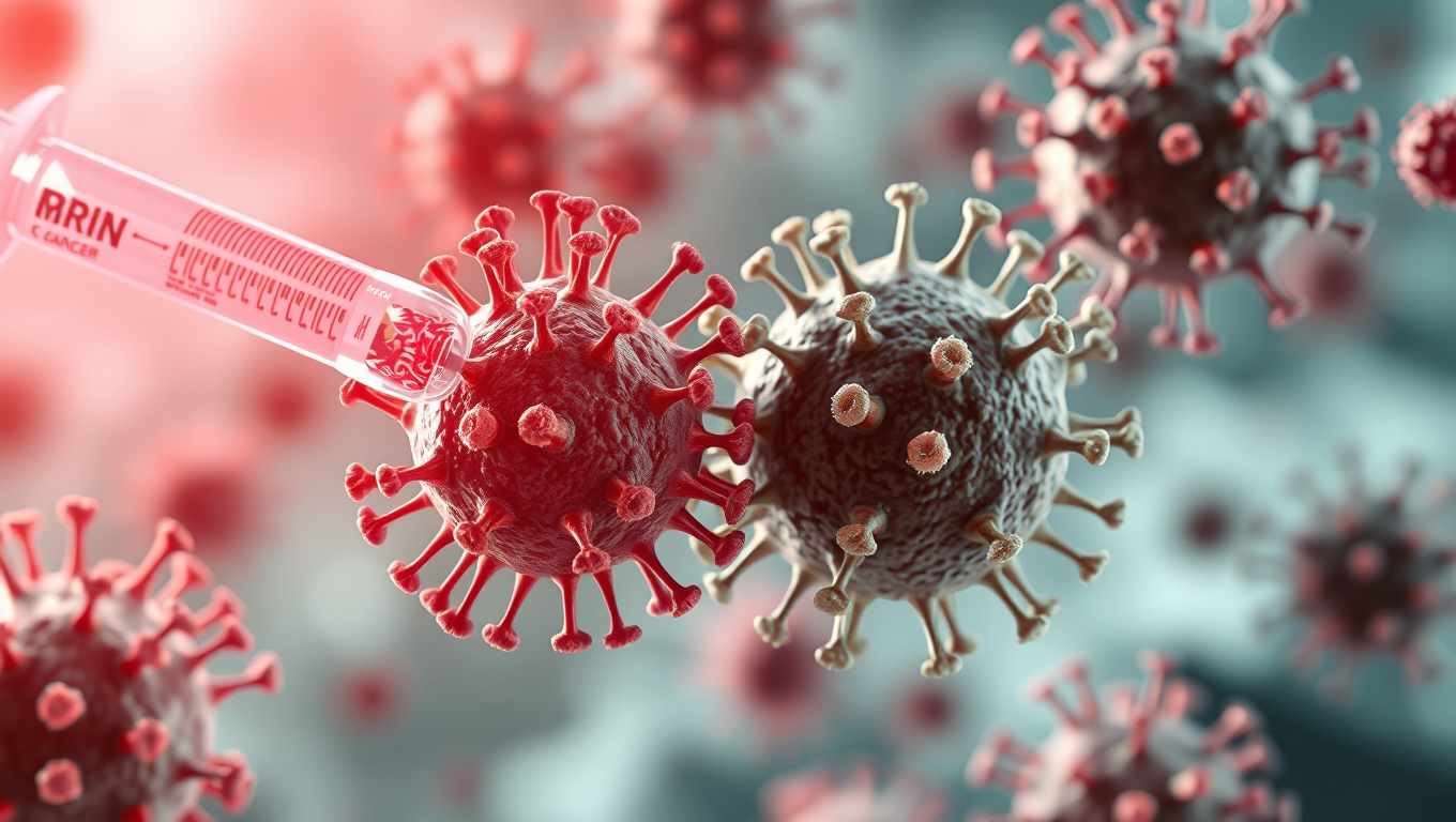
A Breakthrough Cancer Vaccine Shows Promise in Wiping Out Tumors in Mice
A breakthrough mRNA cancer vaccine has shown the ability to supercharge the effects of immunotherapy in mice, sparking hope for a universal “off-the-shelf” treatment that could fight multiple cancers. Unlike traditional vaccines designed to target specific tumor proteins, this approach simply revs up the immune system as if it were fighting a virus. The results were dramatic—when paired with checkpoint inhibitors, tumors shrank, and in some cases, the vaccine alone wiped them out.
Allergy
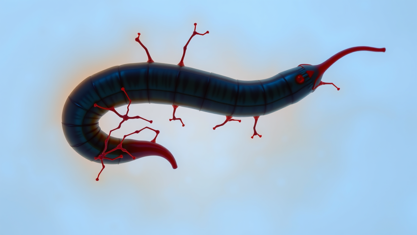
“The Silent Invader: How a Parasitic Worm Evades Detection and What it Can Teach Us About Pain Relief”
Scientists have discovered a parasite that can sneak into your skin without you feeling a thing. The worm, Schistosoma mansoni, has evolved a way to switch off the body’s pain and itch signals, letting it invade undetected. By blocking certain nerve pathways, it avoids triggering the immune system’s alarms. This stealth tactic not only helps the worm survive, but could inspire new kinds of pain treatments and even preventative creams to protect people from infection.
Allergy
Flossing for Vaccines: A New Method to Deliver Immunizations
Scientists have discovered that flossing between your teeth could one day help vaccinate you. By targeting a uniquely permeable gum tissue called the junctional epithelium, this new method stimulates immunity right where many infections enter: the mouth, nose, and lungs. Using dental floss on mice to apply a flu vaccine triggered a robust immune response—better than existing oral approaches and comparable to nasal vaccines, but without the risks. It even worked with mRNA and protein-based vaccines.


A New Horizon for Vision: How Gold Nanoparticles May Restore People’s Sight

Revolutionizing Quantum Communication: Direct Connections Between Multiple Processors

Retiring Abroad Can Be Lonely Business
Harnessing Water Waves: A Breakthrough in Controlling Floating Objects

Household Electricity Three Times More Expensive Than Upcoming ‘Eco-Friendly’ Aviation E-Fuels, Study Reveals

“Unveiling Hidden Patterns: A New Twist on Interference Phenomena”
Reducing Falls Among Elderly Women with Polypharmacy through Exercise Intervention
-
 Detectors1 year ago
A New Horizon for Vision: How Gold Nanoparticles May Restore People’s Sight
-
Cancer1 year ago
Revolutionizing Quantum Communication: Direct Connections Between Multiple Processors
-
Earth & Climate1 year ago
Retiring Abroad Can Be Lonely Business
-
Albert Einstein1 year ago
Harnessing Water Waves: A Breakthrough in Controlling Floating Objects
-
Earth & Climate1 year ago
Household Electricity Three Times More Expensive Than Upcoming ‘Eco-Friendly’ Aviation E-Fuels, Study Reveals
-
Chemistry1 year ago
“Unveiling Hidden Patterns: A New Twist on Interference Phenomena”
-
Diseases and Conditions1 year ago
Reducing Falls Among Elderly Women with Polypharmacy through Exercise Intervention
-
Earth & Climate1 year ago
Predicting Damage from Local Earthquakes in Mexico City