Connect with us
We’re experimenting with AI-generated content to help deliver information faster and more efficiently.
While we try to keep things accurate, this content is part of an ongoing experiment and may not always be reliable.
Please double-check important details — we’re not responsible for how the information is used.

Brain Injury

Unlocking the Secrets of Molecules: How Ion Channels ‘Remember’ and Contribute to Lifelong Learning

Researchers have discovered how an ion channel in the brain’s neurons has a kind of ‘molecular memory’, which contributes to the formation and preservation of lifelong memories. The researchers have identified a specific part of the ion channel at which new drugs for certain genetic diseases could be targeted.
Avatar photo

Published

on

The human brain is capable of incredible feats, from learning new skills to forming lifelong memories. But have you ever wondered how these vital processes work at the molecular level? Researchers at Linköping University in Sweden have made a groundbreaking discovery about an ion channel molecule that plays a crucial role in memory and learning.

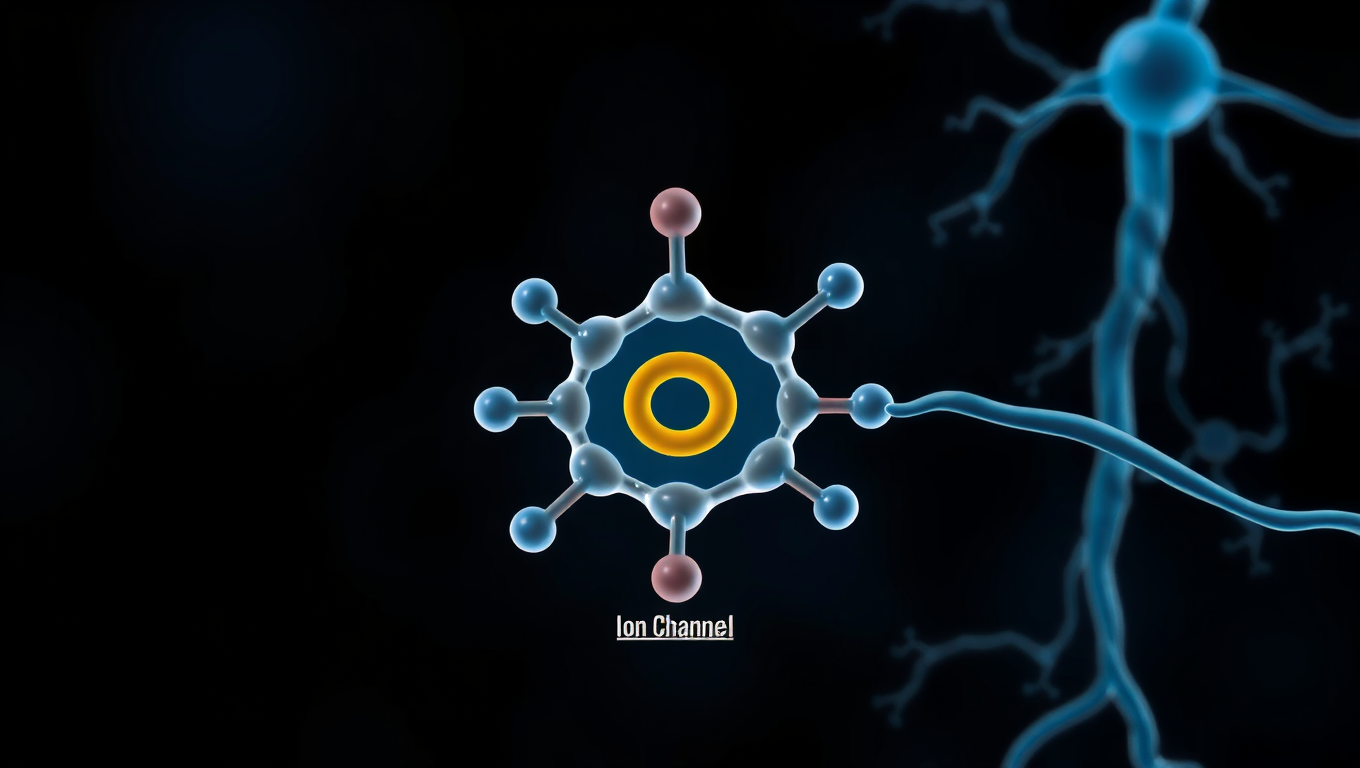
The study focused on a specific type of calcium ion channel called CaV2.1, which is present in the brain’s neurons. These channels are responsible for transmitting signals between neurons through neurotransmitters. However, what’s remarkable about these channels is their ability to “remember” previous electrical signals and adjust their activity accordingly.

“When an electrical signal passes through the neuron, the ion channel opens, setting in motion a process leading to neurotransmitter release,” explains Antonios Pantazis, associate professor at the Department of Biomedical and Clinical Sciences at LiU. “But what we’ve discovered is that prolonged electrical activity can reduce the number of channels that can open, resulting in less transmitter release.”

The researchers found that the ion channel molecule can take almost 200 different shapes depending on the strength and duration of an electrical signal. This complex molecular machine is capable of adapting to changes in its environment, which allows it to “remember” previous signals.

“The channel can then no longer be opened,” says Pantazis. “When hundreds of signals occur over long enough time, they can convert most channels into this ‘declutched memory state’ for several seconds.”

This collective memory in the ion channels can accumulate over time and reduce communication between two neurons. This leads to changes in the receiving neuron that last for hours or days, eventually resulting in much longer-lived changes in the brain.

“In this way, a ‘memory’ that lasts for a few seconds in a single molecule can make a small contribution to a person’s memory that lasts for a lifetime,” Pantazis explains.

The increased knowledge of how calcium ion channels work can contribute to the treatment of certain diseases. Variants of the gene that produces the CaV2.1 channel are linked to rare but serious neurological diseases, which often run in families. By identifying the specific part of the large ion channel that should be targeted when developing new drugs, researchers can take a crucial step towards finding effective treatments for these conditions.

The research has been funded by several organizations, including the Swedish Research Council and the NIH. As scientists continue to uncover the secrets of molecules like the CaV2.1 channel, they may ultimately unlock new possibilities for treating and preventing neurological disorders.

Brain Injury

“Brain Harmony: How Sound Shaping Rewires Your Brain in Real Time”

What happens inside your brain when you hear a steady rhythm or musical tone? According to a new study, your brain doesn’t just hear it — it reorganizes itself in real time.

Avatar photo

Published

on

By

Have you ever wondered what happens inside your brain when you listen to music or a steady rhythm? A groundbreaking study from Aarhus University and the University of Oxford has revealed that your brain doesn’t just hear it – it reorganizes itself in real time. The research, led by Dr. Mattia Rosso and Associate Professor Leonardo Bonetti, introduces a novel neuroimaging method called FREQ-NESS, which maps the brain’s internal organization with high spectral and spatial precision.

Using advanced algorithms, FREQ-NESS disentangles overlapping brain networks based on their dominant frequency, allowing scientists to track how each frequency propagates in space across the brain. This development represents a major advance in neuroscience, enabling researchers to study the brain’s large-scale dynamics more accurately.

The traditional view of brainwaves as fixed stations – alpha, beta, gamma – is being challenged by this research. FREQ-NESS reveals that brain activity is organized through frequency-tuned networks, both internally and externally. This understanding opens new possibilities for basic neuroscience, brain-computer interfaces, and clinical diagnostics.

This study contributes to a growing body of research exploring how the brain’s rhythmic structure shapes perception, attention, and consciousness. Professor Leonardo Bonetti notes, “The brain doesn’t just react – it reconfigures. And now we can see it.” This breakthrough could revolutionize how scientists study brain responses to music and beyond.

A large-scale research program is underway to build on this methodology, supported by an international network of neuroscientists. FREQ-NESS may also pave the way for individualized brain mapping, offering new insights into the intricate harmony of the human brain.

Continue Reading

Artificial Intelligence

Self-Powered Artificial Synapse Revolutionizes Machine Vision

Despite advances in machine vision, processing visual data requires substantial computing resources and energy, limiting deployment in edge devices. Now, researchers from Japan have developed a self-powered artificial synapse that distinguishes colors with high resolution across the visible spectrum, approaching human eye capabilities. The device, which integrates dye-sensitized solar cells, generates its electricity and can perform complex logic operations without additional circuitry, paving the way for capable computer vision systems integrated in everyday devices.

Avatar photo

Published

on

By

The human visual system has long been a source of inspiration for computer vision researchers, who aim to develop machines that can see and understand the world around them with the same level of efficiency and accuracy as humans. While machine vision systems have made significant progress in recent years, they still face major challenges when it comes to processing vast amounts of visual data while consuming minimal power.

One approach to overcoming these hurdles is through neuromorphic computing, which mimics the structure and function of biological neural systems. However, two major challenges persist: achieving color recognition comparable to human vision, and eliminating the need for external power sources to minimize energy consumption.

A recent breakthrough by a research team led by Associate Professor Takashi Ikuno from Tokyo University of Science has addressed these issues with a groundbreaking solution. Their self-powered artificial synapse is capable of distinguishing colors with remarkable precision, making it particularly suitable for edge computing applications where energy efficiency is crucial.

The device integrates two different dye-sensitized solar cells that respond differently to various wavelengths of light, generating its electricity via solar energy conversion. This self-powering capability makes it an attractive solution for industries such as autonomous vehicles, healthcare, and consumer electronics, where visual recognition capabilities are essential but power consumption is limited.

The researchers demonstrated the potential of their device in a physical reservoir computing framework, recognizing different human movements recorded in red, green, and blue with an impressive 82% accuracy. This achievement has significant implications for various industries, including autonomous vehicles, which could utilize these devices to efficiently recognize traffic lights, road signs, and obstacles.

In healthcare, self-powered artificial synapses could power wearable devices that monitor vital signs like blood oxygen levels with minimal battery drain. For consumer electronics, this technology could lead to smartphones and augmented/virtual reality headsets with dramatically improved battery life while maintaining sophisticated visual recognition capabilities.

The realization of low-power machine vision systems with color discrimination capabilities close to those of the human eye is within reach, thanks to this breakthrough research. The potential applications of self-powered artificial synapses are vast, and their impact will be felt across various industries in the years to come.

Continue Reading

Brain Injury

Unlocking the Secret to Weight Loss: Scientists Discover Key Brain Cells that Regulate Semaglutide’s Effects

A specific group of nerve cells in the brain stem appears to control how semaglutide affects appetite and weight — without causing nausea.

Avatar photo

Published

on

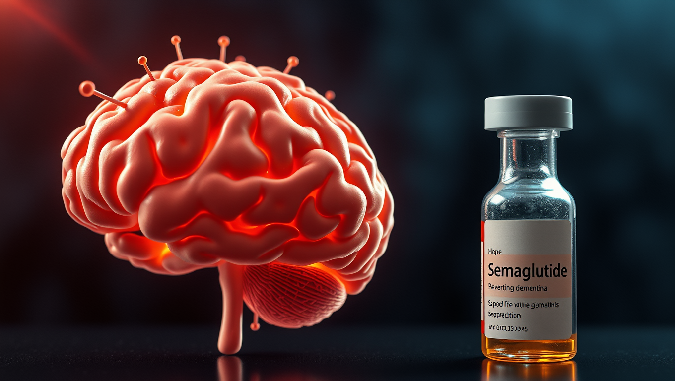
The discovery of a specific group of nerve cells in the brain stem has shed new light on how semaglutide affects appetite and weight loss. Researchers at the University of Gothenburg have made a groundbreaking find that could pave the way for better drugs to treat obesity.

Semaglutide, a GLP-1R agonist, is already well-established as part of the treatment for obesity and type 2 diabetes. However, it can cause side effects such as nausea and muscle loss. The researchers were able to distinguish the nerve cells in the brain that control the beneficial effects of semaglutide from those that contribute to side effects.

In a study published in Cell Metabolism, the researchers worked with mice and tracked which nerve cells were activated by semaglutide. They then stimulated these cells without administering the drug itself. The result was that the mice ate less and lost weight, just as they did when treated with semaglutide. When these nerve cells were killed, the drug’s effect on appetite and fat loss decreased significantly, but side effects such as nausea and muscle loss remained.

“This suggests that these nerve cells control the beneficial effects of semaglutide,” says Júlia Teixidor-Deulofeu, first author of the study. “We have therefore identified a specific group of nerve cells that is necessary for the effects that semaglutide has on weight and appetite, but which does not appear to contribute to any significant extent to side effects such as nausea.”

The identified nerve cells are located in an area of the brain called the dorsal vagal complex. The study provides new knowledge about how semaglutide works in the brain and deeper insight into how the brain stem regulates our energy balance.

“The better we understand this, the greater the opportunity we have to improve them,” says Linda Engström Ruud, researcher and supervisor to PhD students Júlia Teixidor-Deulofeu and Sebastian Blid Sköldheden, who both worked on the project.

This discovery has significant implications for the development of better drugs to treat obesity and could potentially lead to improved treatment options with fewer side effects.

Continue Reading

Trending