Connect with us

Bacteria

Unraveling the Mystery of Post-Treatment Lyme Disease Syndrome: A Breakthrough in Understanding its Causes

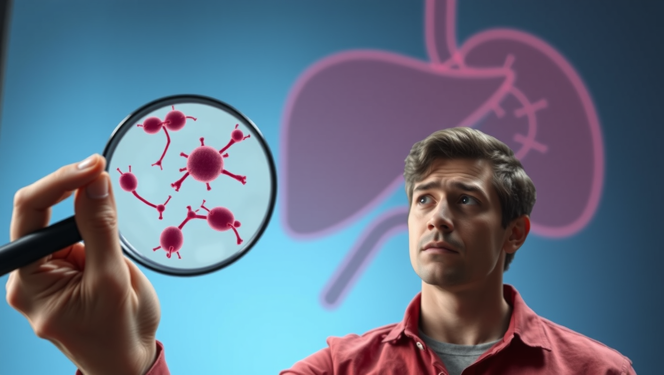
Scientists believe they know what causes the treated infection to mimic chronic illness: the body may be responding to remnants of the bacteria that causes Lyme that tend to pool in the liver and joint fluid.
Avatar photo

Published

on

For years, doctors have been puzzled by the mysterious case of post-treatment Lyme disease syndrome (PTLD), where patients who have received treatment for Lyme disease still experience severe fatigue, cognitive challenges, body pain, and arthritis. A recent study found that 14% of patients who were diagnosed and treated early with antibiotic therapy would still develop PTLD.

Now, Northwestern University scientists believe they have cracked the code to understanding the causes behind this condition. According to Brandon L. Jutras, a bacteriologist leading the research, the body may be responding to remnants of the Borrelia burgdorferi cell wall, which breaks down during treatment yet lingers in the liver.

The key lies in peptidoglycan, a structural feature of virtually all bacterial cells and a common target of antibiotics. Jutras’ team found that while peptidoglycan from other bacteria is rapidly shed after treatment, Lyme disease’s peptidoglycan persists for weeks to months. In humans, pieces of this peptidoglycan were omnipresent in the fluid of patients with Lyme arthritis, even after treatment.

The research suggests that the maladaptive response to these lingering molecules may be behind PTLD. Jutras explained that some patients have a more robust immune response, which could result in a worse disease outcome, while others’ immune systems largely ignore the molecule. This individualized response is likely influenced by genetic factors.

The findings open up new avenues for research and treatment options. Jutras hopes to develop more accurate tests for PTLD patients and refine treatment options when antibiotics have failed. He also proposes neutralizing the inflammatory molecule using monoclonal antibodies to target peptidoglycan for destruction.

With this breakthrough, scientists are one step closer to understanding and effectively treating PTLD, providing relief to millions of people worldwide affected by this debilitating condition.

Bacteria

“Unlocking the Secrets of Wolbachia: How Frisky Flies Could Save Human Lives”

A scientist decided to find out why a bacterial infection makes fruit flies promiscuous. What he discovered could help curb mosquito-borne diseases and manage crop pests.

Avatar photo

Published

on

Timothy Karr, an Arizona State University scientist, has made a groundbreaking discovery that could change the way we combat mosquito-borne diseases and manage crop pests. By studying the effects of Wolbachia, a parasitic bacteria that infects at least two out of every five insect species, on fruit flies, Karr and his team have found that it can make infected females more promiscuous.

Wolbachia’s goal is to spread to more hosts, but it can only pass from an infected mother to her offspring. To improve its chances, it influences its hosts so that infected females lay lots of infected eggs. In fruit flies, Wolbachia makes infected males unable to fertilize uninfected females’ eggs.

Karr and his colleagues set out to study what is happening inside the cells of infected female fruit flies to make them so promiscuous. They found that Wolbachia is perfectly positioned in the regions responsible for mating behavior and decision-making in the brain. Using a protein approach, they compared proteins in infected and uninfected female brains and found over 170 changes.

Three specific proteins were identified as being directly involved in the infection’s effect on mating behavior. By genetically changing their levels in uninfected flies, those flies began acting like the infected ones. Additionally, over 700 Wolbachia proteins were identified in female brains, with two of them interacting with the host fly’s proteins.

These findings have significant implications for managing disease-carrying insects and protecting crops with safer pesticides. Insights from this study might also help protect species like bees that face threats from viruses.

Karr believes that understanding how Wolbachia interacts with its hosts could lead to more lifesaving solutions. He is eager to continue studying the molecular basis of the bacteria’s influence on its hosts, and the team’s success with protein analysis may inspire new studies using this method.

In the words of Karr, “Proteins are where the rubber meets the road.” And it’s a road that could lead to more lifesaving solutions.

Continue Reading

Bacteria

Rivers Around the World Are Being Polluted with Antibiotics from Human Use

Millions of kilometers of rivers around the world are carrying antibiotic pollution at levels high enough to promote drug resistance and harm aquatic life, a new study warns. The study estimated the scale of global river contamination from human antibiotics use. Researchers calculated that about 8,500 tons of antibiotics — nearly one-third of what people consume annually — end up in river systems around the world each year even after in many cases passing through wastewater systems.

Avatar photo

Published

on

Here is the rewritten article:

The world’s rivers are facing an alarming threat: millions of kilometers of waterways are being contaminated with antibiotics from human use. According to a recent study led by McGill University researchers, this pollution has the potential to promote drug resistance and harm aquatic life on a massive scale.

Published in PNAS Nexus, the groundbreaking research is the first to estimate the global scope of river contamination caused by human antibiotic consumption. The team calculated that approximately 8,500 tonnes of antibiotics – about one-third of what people consume annually – end up in river systems worldwide each year, even after passing through wastewater treatment plants.

While individual antibiotic residues might be present at very low concentrations in most rivers, making them difficult to detect, the chronic and cumulative environmental exposure can still pose a risk to human health and aquatic ecosystems. This is particularly concerning for amoxicillin, the world’s most commonly used antibiotic, which was found to be most likely present at risky levels in Southeast Asia.

The region’s rising use of antibiotics combined with limited wastewater treatment has amplified the problem. The study emphasizes that it’s not about discouraging the use of antibiotics – we rely on them for global health treatments. Instead, the findings indicate unintended effects on aquatic environments and antibiotic resistance, which calls for mitigation and management strategies to minimize their implications.

The research used a global model validated by field data from nearly 900 river locations, excluding antibiotics from livestock or pharmaceutical factories, both significant contributors to environmental contamination. The study’s authors suggest that monitoring programs are essential to detect antibiotic or chemical contamination in waterways, especially in areas predicted to be at risk.

In conclusion, the study highlights the critical issue of antibiotic pollution in rivers arising from human consumption alone. While it would likely worsen with contributions from veterinary or industry sources, immediate action is needed to address this pressing concern and protect our planet’s precious aquatic resources.

Continue Reading

Bacteria

Unlocking Efficiency: Researchers Reveal Secrets of Cell Division with Min Proteins

The Min protein system prevents abnormal cell division in bacteria, but is poorly understood. Researchers have uncovered how engineered e.coli bacteria control protein levels for maximum efficiency.

Avatar photo

Published

on

The Min protein system is a complex process that helps bacteria divide evenly and correctly. For decades, scientists have studied this system, but understanding how it works efficiently has been a challenge. Recently, researchers at the University of California San Diego (UCSD) made a groundbreaking discovery that sheds new light on the efficiency of cell division.

The UCSD team developed a way to control Min protein expression levels independently in E. coli cells. This allowed them to observe how different concentrations of Min proteins affect the oscillations between the poles of the cell. The results were surprising: despite varying concentrations, the oscillations remained stable across a wide range, with E. coli producing just the right amount of Min proteins.

This breakthrough is significant because it shows that the Min protein system can efficiently guide division to the correct location without relying on precise control over protein levels. This finding has far-reaching implications for our understanding of cellular organization and function.

The study was published in Nature Physics, a leading scientific journal, and was funded by the National Institutes of Health (NIH). The research team consisted of experts from both physics and chemistry/biochemistry departments at UCSD, highlighting the importance of interdisciplinary collaboration in advancing our knowledge of cellular biology.

Continue Reading

Trending