
While we try to keep things accurate, this content is part of an ongoing experiment and may not always be reliable.
Please double-check important details — we’re not responsible for how the information is used.
Amyotrophic Lateral Sclerosis
Precision Medicine Breakthrough: Researchers Develop Tool to Manipulate Mitochondrial DNA
Many mitochondrial diseases have been difficult to study and treat due to the inherent challenges in accessing mitochondrial DNA (mtDNA). Now, researchers have optimized mitochondrial-targeted compounds that can selectively modify the ratio of normal versus mutant mtDNA in patient-derived stem cells. This technology enables the creation of research models with varying mutation loads and demonstrates potential as a therapeutic strategy for reducing mutant mtDNA in patients, offering hope for mitochondrial disease treatment.


Amyotrophic Lateral Sclerosis
“Reviving Memories: Gene Therapy Shows Promise in Reversing Alzheimer’s Disease in Mice”
UC San Diego scientists have created a gene therapy that goes beyond masking Alzheimer’s symptoms—it may actually restore brain function. In mice, the treatment protected memory and altered diseased brain cells to behave more like healthy ones.

Alzheimer's
Different Versions of APOE Protein Alter Microglia Function in Alzheimer’s Disease
A new study suggests how APOE2 is protective while APOE4 increases disease risk by regulating the brain’s immune cells.

Alzheimer's
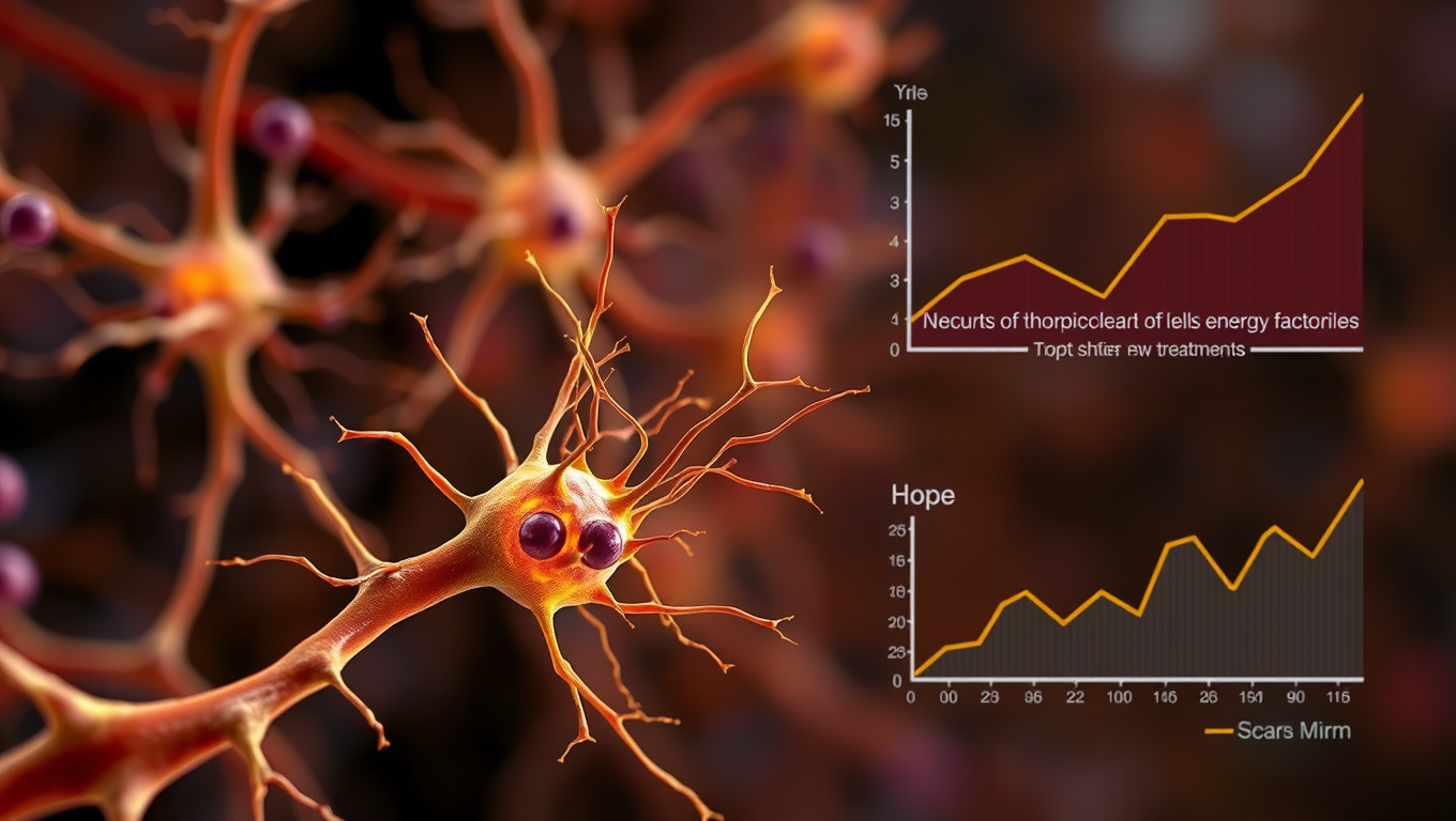
Breaking Ground in ALS Research: Uncovering Early Signs of Disease and New Treatment Targets
Using the gene scissors CRISPR and stem cells, researchers have managed to identify a common denominator for different gene mutations that all cause the neurological disease ALS. The research shows that ALS-linked dysfunction occurs in the energy factories of nerve cells, the mitochondria, before the cells show other signs of disease, which was not previously known.

A New Horizon for Vision: How Gold Nanoparticles May Restore People’s Sight

Revolutionizing Quantum Communication: Direct Connections Between Multiple Processors

Retiring Abroad Can Be Lonely Business
Harnessing Water Waves: A Breakthrough in Controlling Floating Objects

Household Electricity Three Times More Expensive Than Upcoming ‘Eco-Friendly’ Aviation E-Fuels, Study Reveals

“Unveiling Hidden Patterns: A New Twist on Interference Phenomena”
Reducing Falls Among Elderly Women with Polypharmacy through Exercise Intervention
-
 Detectors1 year ago
A New Horizon for Vision: How Gold Nanoparticles May Restore People’s Sight
-
Cancer1 year ago
Revolutionizing Quantum Communication: Direct Connections Between Multiple Processors
-
Earth & Climate1 year ago
Retiring Abroad Can Be Lonely Business
-
Albert Einstein1 year ago
Harnessing Water Waves: A Breakthrough in Controlling Floating Objects
-
Earth & Climate1 year ago
Household Electricity Three Times More Expensive Than Upcoming ‘Eco-Friendly’ Aviation E-Fuels, Study Reveals
-
Chemistry1 year ago
“Unveiling Hidden Patterns: A New Twist on Interference Phenomena”
-
Diseases and Conditions1 year ago
Reducing Falls Among Elderly Women with Polypharmacy through Exercise Intervention
-
Agriculture and Food1 year ago
“A Sustainable Solution: Researchers Create Hybrid Cheese with 25% Pea Protein”


