
While we try to keep things accurate, this content is part of an ongoing experiment and may not always be reliable.
Please double-check important details — we’re not responsible for how the information is used.
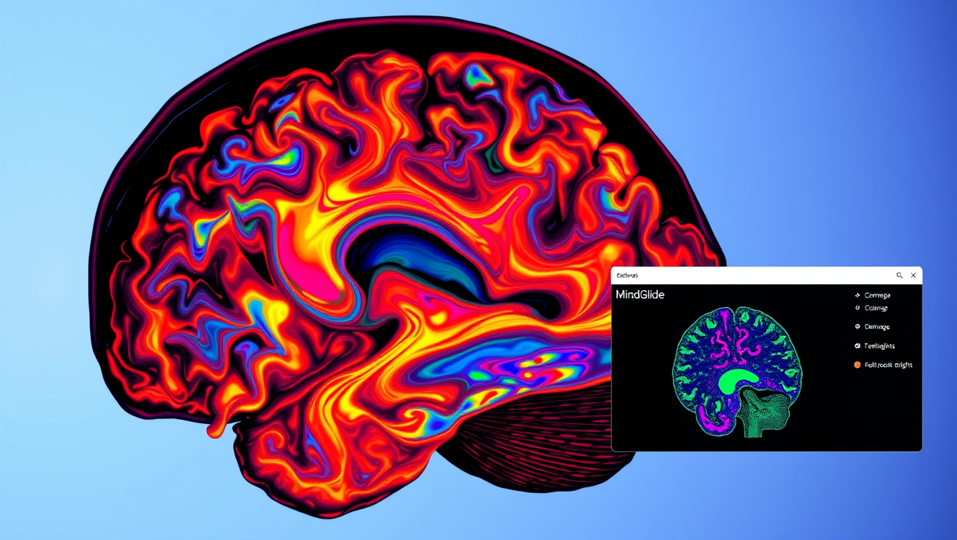
Brain Injury
“Revolutionizing Multiple Sclerosis Treatment with AI: UCL Researchers’ Breakthrough Tool, MindGlide”
A new artificial intelligence (AI) tool that can help interpret and assess how well treatments are working for patients with multiple sclerosis (MS) has been developed.


Alzheimer's
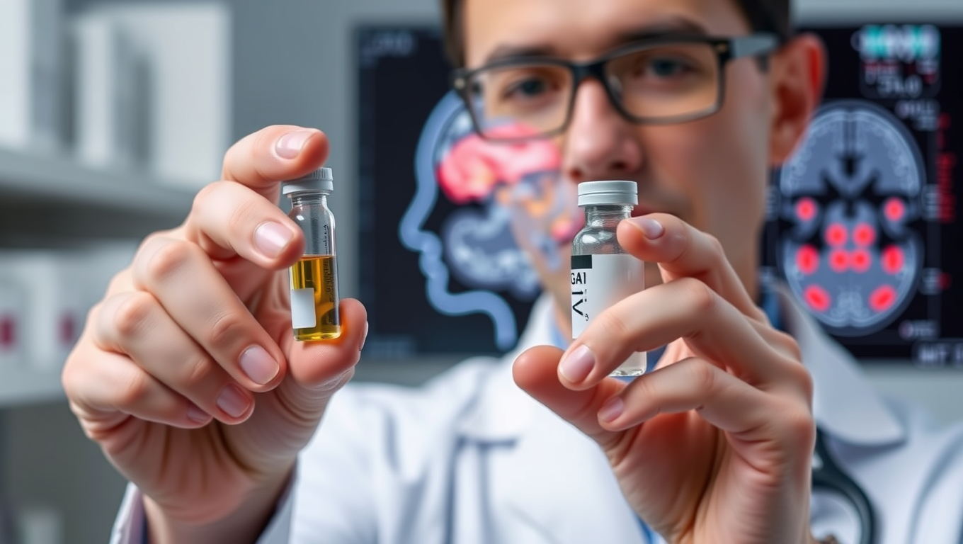
Rewinding Stroke Damage and Beyond: The Promise of GAI-17
Stroke kills millions, but Osaka researchers have unveiled GAI-17, a drug that halts toxic GAPDH clumping, slashes brain damage and paralysis in mice—even when given six hours post-stroke—and shows no major side effects, hinting at a single therapy that could also tackle Alzheimer’s and other tough neurological disorders.
Brain Injury
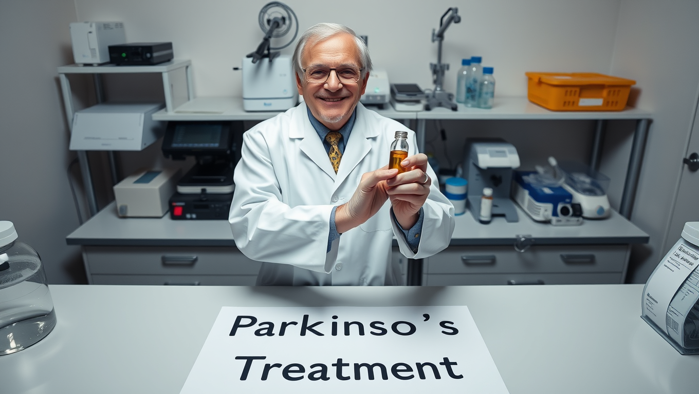
Scientists Edge Closer to Reversing Parkinson’s Symptoms — A Breakthrough for Humans?
Scientists at the University of Sydney have uncovered a malfunctioning version of the SOD1 protein that clumps inside brain cells and fuels Parkinson’s disease. In mouse models, restoring the protein’s function with a targeted copper supplement dramatically rescued movement, hinting at a future therapy that could slow or halt the disease in people.
Alzheimer's
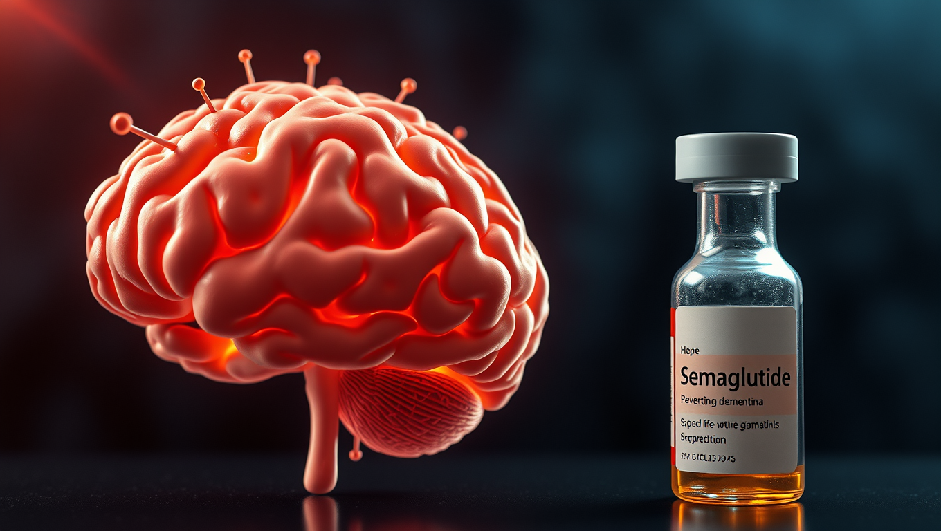
Groundbreaking Study Suggests Link Between Semaglutide and Lower Dementia Risk in Type 2 Diabetes Patients
A blockbuster diabetes and weight-loss drug might be doing more than controlling blood sugar—it could also be protecting the brain. Researchers at Case Western Reserve University found that people with type 2 diabetes who took semaglutide (the active ingredient in Ozempic and Wegovy) had a significantly lower risk of developing dementia. The benefit was especially strong in women and older adults.

A New Horizon for Vision: How Gold Nanoparticles May Restore People’s Sight

Retiring Abroad Can Be Lonely Business

Revolutionizing Quantum Communication: Direct Connections Between Multiple Processors
Harnessing Water Waves: A Breakthrough in Controlling Floating Objects

“Unveiling Hidden Patterns: A New Twist on Interference Phenomena”

Household Electricity Three Times More Expensive Than Upcoming ‘Eco-Friendly’ Aviation E-Fuels, Study Reveals
Reducing Falls Among Elderly Women with Polypharmacy through Exercise Intervention
-
 Detectors12 months ago
A New Horizon for Vision: How Gold Nanoparticles May Restore People’s Sight
-
Earth & Climate1 year ago
Retiring Abroad Can Be Lonely Business
-
Cancer1 year ago
Revolutionizing Quantum Communication: Direct Connections Between Multiple Processors
-
Albert Einstein1 year ago
Harnessing Water Waves: A Breakthrough in Controlling Floating Objects
-
Chemistry1 year ago
“Unveiling Hidden Patterns: A New Twist on Interference Phenomena”
-
Earth & Climate1 year ago
Household Electricity Three Times More Expensive Than Upcoming ‘Eco-Friendly’ Aviation E-Fuels, Study Reveals
-
Diseases and Conditions1 year ago
Reducing Falls Among Elderly Women with Polypharmacy through Exercise Intervention
-
Agriculture and Food1 year ago
“A Sustainable Solution: Researchers Create Hybrid Cheese with 25% Pea Protein”


